

Electroporation of the vasculature and the lung
- PMID: 14683590
- PMCID: PMC4403238
- DOI: 10.1089/104454903322625000
Electroporation of the vasculature and the lung
Abstract
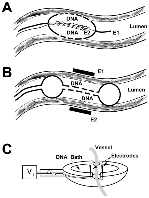
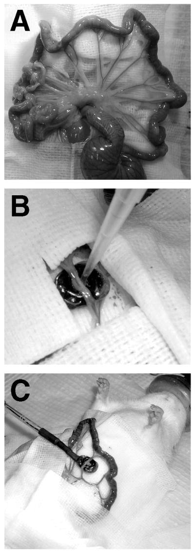
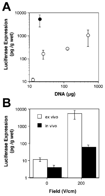
Electroporation has proven to be a highly effective technique for the in vivo delivery of genes to a number of solid tissues. In most of the reported methods, DNA is injected into the target tissue and electrodes are placed directly on or in the tissue for application of the electric field. While this works well for solid tissues, there are many tissues and organs that are not amenable to such an approach. In this review I will focus on the development of electroporation protocols for two such tissues: the vasculature and the lung. Several methods for in vivo electroporation of the vasculature have been developed in recent years that deliver DNA to vessel segments from either the inside or outside of the vessel. The advantages and disadvantages of each are discussed, as are the applications for which they have been used. In more recent work, our laboratory has developed a novel method to deliver genes to the rodent lung that results in high level, uniform, gene expression throughout all cell types of the lung. Most importantly, this technique is safe, and causes no inflammatory response or alterations in normal physiology of the organs. Taken together, these studies demonstrate the utility of electroporation for gene transfer to non injectible tissues.
Figures
References
-
- AIHARA H, MIYAZAKI J. Gene transfer into muscle by electroporation in vivo. Nat Biotechnol. 1998;16:867–870. - PubMed
-
- AUSUBEL FM, BRENT R, KINGSTON RE, MOORE DD, SEIDMAN JG, SMITH JA, STRUHL K, editors. Short protocols in molecular biology. New York: John Wiley & Sons; 1999.
-
- BARRON L, UYECHI L, SZOKA FC. Cationic lipids are essential for gene delivery mediated by intravenous administration of lipoplexes. Gene Ther. 1999;6:1179–1183. - PubMed
-
- BEBOK Z, ABAI AM, DONG JY, KING SA, KIRK KL, BERTA G, HUGHES BW, KRAFT AS, BURGESS SW, SHAW W, et al. Efficiency of plasmid delivery and expression after lipid-mediated gene transfer to human cells in vitro. J Pharmacol Exp Ther. 1996;279:1462–1469. - PubMed
Publication types
MeSH terms
Substances
Grants and funding
LinkOut - more resources
Full Text Sources
Other Literature Sources
