

Changes in MRI signal intensity during hypercapnic challenge under conscious and anesthetized conditions
- PMID: 14684202
- PMCID: PMC2962949
- DOI: 10.1016/s0730-725x(03)00204-2
Changes in MRI signal intensity during hypercapnic challenge under conscious and anesthetized conditions
Abstract
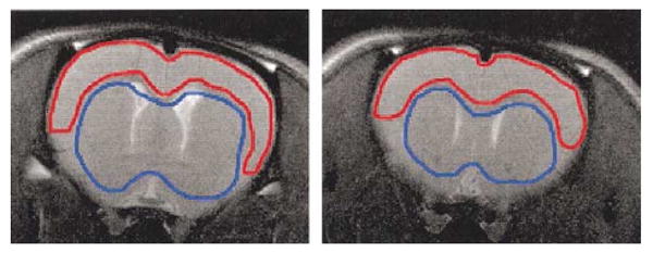
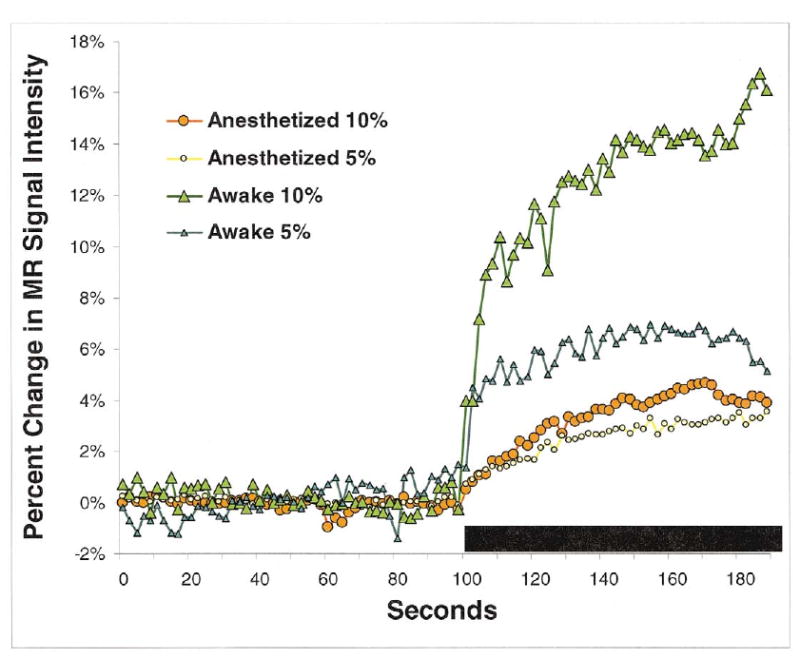
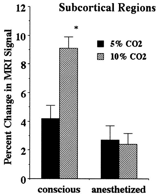
Most functional magnetic resonance imaging (fMRI) studies in animals are conducted under anesthesia to minimize motion artifacts. However, methods and techniques have been developed recently for imaging fully conscious rats. Functional MRI studies on conscious animals report enhanced BOLD signal changes as compared to the anesthetized condition. In this study, rats were exposed to different concentrations of carbon dioxide (CO(2)) while conscious and anesthetized to test whether cerebrovascular reactivity may be contributing to these enhanced BOLD signal changes. Hypercapnia produced significantly greater increases in MRI signal intensity in fully conscious animals (6.7-13.3% changes) as when anesthetized with 1% isoflurane (3.2-4.9% changes). In addition, the response to hypercapnia was more immediate in the conscious condition (< 30s) with signal risetimes twice as fast as in the anesthetized state (60s). Both cortical and subcortical brain regions showed a robust, dose- dependent increase in MRI signal intensity with hypercapnic challenge while the animals were conscious but little or no change when anesthetized. Baseline variations in MRI signal were higher while animals were conscious but this was off set by greater signal intensity changes leading to a greater contrast-to-noise ratio, 13.1 in conscious animals, as compared to 8.0 in the anesthetized condition. In summary, cerebral vasculature appears to be more sensitive to hypercapnic challenge in the conscious condition resulting in enhanced T2* MRI signal intensity and the potential for better BOLD signal changes during functional imaging.
Figures

References
-
- Hajnal JV, Myers R, Oatridge A, Schwieso JE, Young IR, Bydder GM. Artifacts due to stimulus correlated motion in functional imaging of the brain. Magn Reson Med. 1994;31(3):283–91. - PubMed
-
- Bim RM, Bandettini PA, Cox RW, Jesmanowicz A, Shaker R. Magnetic field changes in the human brain due to swallowing or speaking. Magn Reson Med. 1998;40(1):55–60. - PubMed
-
- Ueki M, Mies G, Hossmann KA. Effect of α-chloralose, halothane, pentobarbital and nitrous oxide anesthesia on metabolic coupling in somatosensory cortex of rat. Acta Anaesthesiol Scand. 1992;36(4):318–22. - PubMed
-
- Bonvento G, Seylaz J, Lacombe P. Widespread attenuation of the cerebrovascular reactivity to hypercapnia following inhibition of nitric oxide synthase in the conscious rat. J Cereb Blood Flow Metab. 1994 - PubMed
Publication types
MeSH terms
Substances
Grants and funding
LinkOut - more resources
Full Text Sources
Medical
