Defective mucosal T cell death is sustainably reverted by infliximab in a caspase dependent pathway in Crohn's disease
- PMID: 14684579
- PMCID: PMC1773915
- DOI: 10.1136/gut.53.1.70
Defective mucosal T cell death is sustainably reverted by infliximab in a caspase dependent pathway in Crohn's disease
Abstract
Background and aims: To verify whether targeting defective mucosal T cell death underlies the sustained therapeutic benefit of infliximab in Crohn's disease, we explored its in vivo proapoptotic effect after 10 weeks of treatment, and its in vitro killing activity on lamina propria T cells (LPT) and peripheral blood T cells (PBT), both isolated from Crohn's disease patients.
Methods: Endoscopic intestinal biopsies were collected from 10 Crohn's disease patients (six steroid refractory and four fistulising) before and after three consecutive infusions of infliximab, administered at week 0, 2, and 6 in a single intravenous dose (5 mg/kg), and from 10 subjects who proved to have functional diarrhoea. Apoptosis was determined in vivo by TUNEL assay, and in vitro by fluorescein isothiocyanate-annexin V/propidium iodide staining on LPT and PBT from Crohn's disease patients cultured with infliximab. The effect of the broad caspase inhibitor Z-VAD-FMK and the neutralising anti-Fas antibody ZB4 was tested in vitro on LPT and PBT treated with infliximab. Caspase-3 activity was determined by immunoblotting.
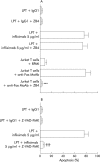
Results: In Crohn's disease patients, infliximab treatment induced a sustained LPT apoptosis, still evident four weeks after the last infusion. In vitro infliximab induced death of LPT from Crohn's disease patients occurred via apoptosis rather than necrosis. LPT showed a higher susceptibility to infliximab induced apoptosis than PBT in Crohn's disease patients. The signalling pathway underlying the restoration of infliximab induced LPT apoptosis occurred via the caspase pathway but not Fas-Fas ligand interaction in Crohn's disease.
Conclusions: These findings demonstrate that apoptosis is the major mechanism by which infliximab exerts its killing activity on LPT in Crohn's disease. The sustained LPT proapoptotic action of infliximab, which extends far beyond its circulating half life, may be responsible for the sustained remission induced in Crohn's disease patients by infliximab retreatment.
Figures


Comment in
-
Differential modulation of p38 mitogen activated protein kinase and STAT3 signalling pathways by infliximab and etanercept in intestinal T cells from patients with Crohn's disease.Gut. 2005 Feb;54(2):314-5; author reply 316-6. Gut. 2005. PMID: 15647208 Free PMC article. No abstract available.
Similar articles
-
Infliximab but not etanercept induces apoptosis in lamina propria T-lymphocytes from patients with Crohn's disease.Gastroenterology. 2003 Jun;124(7):1774-85. doi: 10.1016/s0016-5085(03)00382-2. Gastroenterology. 2003. PMID: 12806611
-
Prediction of antitumour necrosis factor clinical efficacy by real-time visualisation of apoptosis in patients with Crohn's disease.Gut. 2007 Apr;56(4):509-17. doi: 10.1136/gut.2006.105379. Epub 2006 Nov 2. Gut. 2007. PMID: 17082252 Free PMC article. Clinical Trial.
-
Increased production of granulocyte-macrophage colony-stimulating factor in Crohn's disease--a possible target for infliximab treatment.Eur J Gastroenterol Hepatol. 2004 Jul;16(7):649-55. doi: 10.1097/01.meg.0000108344.41221.8b. Eur J Gastroenterol Hepatol. 2004. PMID: 15201577
-
Review article: efficacy of infliximab in Crohn's disease--induction and maintenance of remission.Aliment Pharmacol Ther. 1999 Sep;13 Suppl 4:9-15; discussion 38. doi: 10.1046/j.1365-2036.1999.00025.x. Aliment Pharmacol Ther. 1999. PMID: 10597334 Review.
-
Infliximab: an updated review of its use in Crohn's disease and rheumatoid arthritis.BioDrugs. 2002;16(2):111-48. doi: 10.2165/00063030-200216020-00005. BioDrugs. 2002. PMID: 11985485 Review.
Cited by
-
Cellular and humoral mechanisms involved in the control of tuberculosis.Clin Dev Immunol. 2012;2012:193923. doi: 10.1155/2012/193923. Epub 2012 May 17. Clin Dev Immunol. 2012. PMID: 22666281 Free PMC article. Review.
-
Effects of Anti-Cytokine Antibodies on Gut Barrier Function.Mediators Inflamm. 2019 Nov 12;2019:7028253. doi: 10.1155/2019/7028253. eCollection 2019. Mediators Inflamm. 2019. PMID: 31780866 Free PMC article. Review.
-
Genetically determined high activity of IL-12 and IL-18 in ulcerative colitis and TLR5 in Crohns disease were associated with non-response to anti-TNF therapy.Pharmacogenomics J. 2018 Jan;18(1):87-97. doi: 10.1038/tpj.2016.84. Epub 2017 Jan 31. Pharmacogenomics J. 2018. PMID: 28139755
-
Anti-tumour necrosis factor therapy enhances mucosal healing through down-regulation of interleukin-21 expression and T helper type 17 cell infiltration in Crohn's disease.Clin Exp Immunol. 2013 Jul;173(1):102-11. doi: 10.1111/cei.12084. Clin Exp Immunol. 2013. PMID: 23607532 Free PMC article.
-
Ex vivo immunosuppressive effects of mesenchymal stem cells on Crohn's disease mucosal T cells are largely dependent on indoleamine 2,3-dioxygenase activity and cell-cell contact.Stem Cell Res Ther. 2015 Jul 24;6(1):137. doi: 10.1186/s13287-015-0122-1. Stem Cell Res Ther. 2015. PMID: 26206376 Free PMC article.
References
-
- Fuss IJ, Marth T, Neurath MF, et al. Anti-interleukin 12 treatment regulates apoptosis of Th1 T cells in experimental colitis in mice. Gastroenterology 1999;117:1078–88. - PubMed
-
- Atreya R, Mudter J, Finotto S, et al. Blockade of interleukin 6 trans signaling suppresses T-cell resistance against apoptosis in chronic intestinal inflammation: evidence in Crohn disease and experimental colitis in vivo. Nat Med 2000;6:583–8. - PubMed
-
- Lügering A, Schmidt M, Lügering N, et al. Infliximab induces apoptosis in monocytes from patients with chronic active Crohn’s disease by using a caspase-dependent pathway. Gastroenterology 2001;121:1145–57. - PubMed
MeSH terms
Substances
LinkOut - more resources
Full Text Sources
Medical
Research Materials
Miscellaneous
