. 2004 Jan;134(1):307-19.
doi: 10.1104/pp.103.028712. Epub 2003 Dec 18.

Biocontrol of Bacillus subtilis against infection of Arabidopsis roots by Pseudomonas syringae is facilitated by biofilm formation and surfactin production

Affiliations

Biocontrol of Bacillus subtilis against infection of Arabidopsis roots by Pseudomonas syringae is facilitated by biofilm formation and surfactin production

Harsh Pal Bais et al. Plant Physiol. 2004 Jan.

Abstract

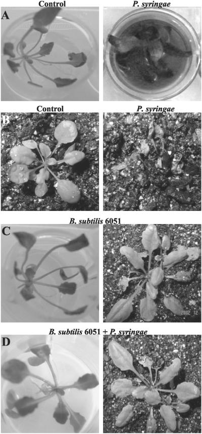
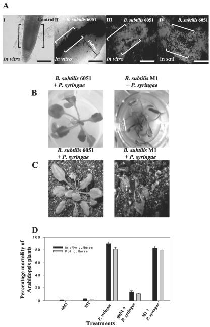
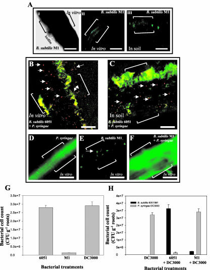
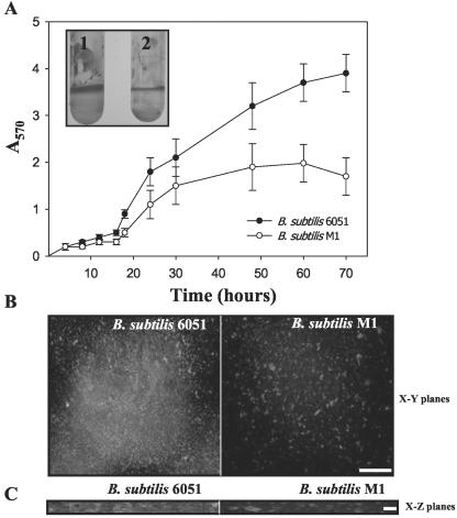
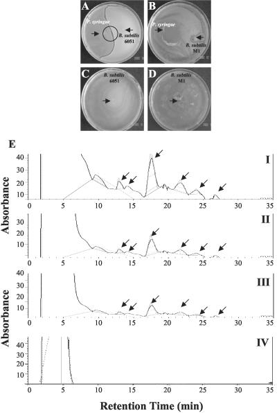
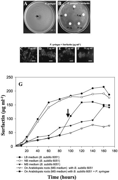
Relatively little is known about the exact mechanisms used by Bacillus subtilis in its behavior as a biocontrol agent on plants. Here, we report the development of a sensitive plant infection model demonstrating that the bacterial pathogen Pseudomonas syringae pv tomato DC3000 is capable of infecting Arabidopsis roots both in vitro and in soil. Using this infection model, we demonstrated the biocontrol ability of a wild-type B. subtilis strain 6051 against P. syringae. Arabidopsis root surfaces treated with B. subtilis were analyzed with confocal scanning laser microscopy to reveal a three-dimensional B. subtilis biofilm. It is known that formation of biofilms by B. subtilis is a complex process that includes secretion of surfactin, a lipopeptide antimicrobial agent. To determine the role of surfactin in biocontrol by B. subtilis, we tested a mutant strain, M1, with a deletion in a surfactin synthase gene and, thus, deficient in surfactin production. B. subtilis M1 was ineffective as a biocontrol agent against P. syringae infectivity in Arabidopsis and also failed to form robust biofilms on either roots or inert surfaces. The antibacterial activity of surfactin against P. syringae was determined in both broth and agar cultures and also by live-dead staining methods. Although the minimum inhibitory concentrations determined were relatively high (25 microg mL(-1)), the levels of the lipopeptide in roots colonized by B. subtilis are likely to be sufficient to kill P. syringae. Our results collectively indicate that upon root colonization, B. subtilis 6051 forms a stable, extensive biofilm and secretes surfactin, which act together to protect plants against attack by pathogenic bacteria.


Figures

Figure 1.
Figure 1.
A, Pathogenicity of the P. syringae strain DC3000 against Arabidopsis in vitro. P. syringae was infiltrated into the liquid Murashige and Skoog medium of in vitro-grown plants, and plant mortality was recorded 7 d postinoculation. B, Pathogenicity of P. syringae strain DC3000 against Arabidopsis in soil. Bacteria were added to sterile soil of plants and plant mortality was again recorded 7 d postinoculation. C, In vitro- and soil-grown Arabidopsis plants cocultivated with B. subtilis 6051. Arabidopsis treated with B. subtilis 6051 alone did not exhibit any plant mortality under in vitro and soil conditions. D, The biocontrol ability of B. subtilis 6051 was checked by inoculating a known concentration of bacterial inoculum into the liquid medium of in vitro- and soil-grown Arabidopsis plants. Arabidopsis plants cocultivated with B. subtilis 6051 and subsequently infected with P. syringae DC3000 under in vitro and soil conditions. P. syringae DC3000 was added on the 4 d post-cocultivation of B. subtilis with Arabidopsis roots. Pretreatment with B. subtilis 6051 reduced plant mortality by approximately 70%.
Figure 2.
Figure 2.
A, Phase contrast microscopy and CSLM of Arabidopsis roots cocultivated with B. subtilis 6051 under in vitro and soil conditions for 4 d showed mature biofilm formation by B. subtilis 6051. I, Phase contrast image of untreated roots (the control). II, Roots treated with the B. subtilis 6051 strain under in vitro conditions; note the marked area indicating a phase-bright material suggestive of biofilm surrounding the roots. III, CSLM image of roots treated with B. subtilis 6051 strain under in vitro conditions. IV, CSLM image of roots visualized after cocultivation with B. subtilis 6051 strain under soil conditions. Double brackets in I to IV indicate the roots. Bars = 50 μm. A live-dead BacLight bacterial viability kit was used to detect the live bacteria. I to IV depict a section of the root (from the root tip to the central elongation zone). B, Interaction of Arabidopsis-B. subtilis 6051 and Arabidopsis-B. subtilis M1 cocultures with P. syringae was checked by inoculating a known concentration of P. syringae inoculum under in vitro conditions. B, Lack of biocontrol ability in B. subtilis M1. C, P. syringae was added on the 4th d post-cocultivation of the two B. subtilis strains with Arabidopsis in soil. D, Plant mortality was quantified for each treatment by scoring the percentage of dead plants over the percentage of live ones to measure biocontrol under both in vitro and soil conditions. Bars = one se. Two-way ANOVA for plant mortality: Ftreatment, 12.12; degrees of freedom, 1.26; and P < 0.001.
Figure 3.
Figure 3.
A, Phase-contrast and CSLM of Arabidopsis roots cocultured with the B. subtilis M1 mutant under in vitro and soil conditions showing poor biofilm formation by the B. subtilis M1 mutant. I, Phase contrast image of roots treated with B. subtilis M1; note the marked area indicating a small region of phase-bright material suggestive of poor biofilm surrounding the roots. II, CSLM image of roots treated with B. subtilis M1 under in vitro conditions. III, CSLM image of Arabidopsis roots cocultivated with B. subtilis M1 under soil conditions. Double brackets in I to III indicate the roots. Bars = 50 μm. I to III depict a section of the root (from the root tip to the central elongation zone). B, CSLM of Arabidopsis roots grown in vitro, cocultivated with B. subtilis 6051, and subsequently infected with P. syringae. C, CSLM of Arabidopsis roots grown under soil conditions, cocultivated with B. subtilis 6051, and subsequently infected with P. syringae. A live-dead BacLight bacterial viability kit was used to detect most of the live bacteria. Bacteria on the root surface were stained with propidium iodide and SYTO9 (See “Materials and Methods”) to visualize the polysaccharides. Both the above treatments show intact biofilm formation by wild-type B. subtilis 6051 as represented by green communities. (Note multiple arrows indicating dead bacteria, most likely P. syringae, in red; brackets in the panel indicate the roots). Bars (B and C) = 20 μm. D, CSLM of P. syringae alone infecting Arabidopsis roots. E, CSLM of Arabidopsis roots grown in vitro and cocultured with the B. subtilis M1 strain. Note the arrow indicating poor colonization as represented by green communities in patches. Bars (D and E) = 50 μm. F, CSLM of Arabidopsis roots grown in vitro, cocultured with B. subtilis M1, and subsequently infected with P. syringae. Brackets in the panels indicate the roots. G and H, Quantification of bacterial cell counts on root surfaces of Arabidopsis on the 4th d postinoculation with B. subtilis 6051, M1, and P. syringae under lone and mixed treatments. Specific antibiotic selection with B. subtilis M1 (5 μg mL-1 chloramphenicol) and P. syringae DC3000 (100 μg mL-1 rifampicin) was used for selective plating of the two bacteria for colony-forming units (cfu) counts. Values show the quantitative amount of roots with average (mean ± sd; n = 5) bacterial counts after inoculation of approximately 2.5 × 10⁸ cfu mL-1; five plants of each species were used per treatment.
Figure 4.
Figure 4.
A, OD readings from microtiter plate assays of biofilm formation by B. subtilis 6051 wild-type and M1 mutant strains. OD570 of solubilized CV from microtiter assays over time for the two tested B. subtilis strains; inset shows the solubilized CV in polypropylene tubes depicting adherent biofilms: 1, B. subtilis 6051; and 2, B. subtilis M1 (values are mean ± sd, n = 5). B, CSLM visualization of wild-type 6051 and M1 mutant strains of B. subtilis grown on glass coverslips. C, An artist's rendering of the three-dimensional structure of biofilm formed by wild-type (6051) and M1 mutant strains of B. subtilis on x-z planes as visualized by CSLM. Bars = 5 μm.
Figure 5.
Figure 5.
A and B, In situ challenge of B. subtilis 6051 and P. syringae bacterial cultures. Bacterial cultures were inoculated with a sterile toothpick on each one-half of the petri plate. Inhibition zones were visualized and photographed 48 h postinoculation. Plates indicate formation of surface film in NB agar medium (see “Materials and Methods”). A, B. subtilis 6051 shows a clear and distinct inhibition zone repelling P. syringae, independent of the medium used (note the arrow indicating the bacterial inocula). B, B. subtilis M1 was out-competed by P. syringae, a result independent of medium used (note the arrows indicating the bacterial inocula). C and D, Lone B. subtilis 6051 and M1 growth plates on NB agar medium (note the arrows indicating bacterial inocula). E, HPLC spectrograms for surfactin production. Six peaks were used to quantify surfactin content in B. subtilis. (Note the arrows indicating the six different isoforms of surfactin). I, Standard surfactin. II, Surfactin content in B. subtilis 6051 broth cultures. III, Surfactin content isolated from the interface of the inhibition zone observed during competition experiment between B. subtilis 6051 and P. syringae. IV, No surfactin was found from the B. subtilis M1 mutant.
Figure 6.
Figure 6.
A and B, Antibacterial activity of 1 to 25 μg mL-1 surfactin on the growth of P. syringae. Standard surfactin was applied to the filter discs. A bacterial inoculum (approximately 2 × 10⁸ cells mL-1) of P. syringae was plated and spread on the petri dish, and the radial inhibition was observed on an hourly basis. A filter disc treated with only solvent (2.5% [v/v] dimethyl sulfoxide) was used as a negative control. A, Radial and efficient growth of P. syringae on NB agar plates in the absence of surfactin. B, Radial growth inhibition of P. syringae in the presence of different concentrations of surfactin. C to F, Fluorescence microscopic visualization of live-dead staining to show bactericidal activity of surfactin against P. syringae in the titer plate assay. Bacterial suspensions treated with sub-MIC (0-5 μg mL-1), MIC (25 μg mL-1), and double MIC levels (50 μg mL-1) of surfactin were stained with propidium iodide and SYTO9 (see “Materials and Methods”) to visualize the polysaccharides and nuclei, respectively. The green fluorescence in C and D depict live material surrounding/inside the bacterial colony. Red fluorescence shows dead cells (E and F). Scale bars = 20 μm. G, Surfactin profiles under different media conditions (LB, NB, and Murashige and Skoog) and on the root surface of Arabidopsis grown alone, cocultured with B. subtilis strains, and infected with P. syringae. (Note the arrow indicating 96 h, the time of addition of P. syringae to the Arabidopsis-B. subtilis 6051 coculture). Arabidopsis roots were weighed (50 mg fresh weight) and extracted for surfactin analysis. (Values are mean ± sd, n = 5).

References

    1. Amellal N, Burtin G, Bartoli F, Heulin T (1998) Colonization of wheat roots by an exopolysaccharide-producing pantoea agglomerans strain and its effect on rhizosphere soil aggregation. Appl Environ Microbiol 64: 3740-3747 - PMC - PubMed
    1. Anagnostopolous C, Spizizen J (1961) Requirements for transformation in Bacillus subtilis. J Bacteriol 81: 741-746 - PMC - PubMed
    1. Asaka O, Shoda M (1996) Biocontrol of Rhizoctonia solani damping off of Tomato with Bacillus subtilis RB14. Appl Environ Microbiol 62: 4081-4085 - PMC - PubMed
    1. Bashan Y, Bashan LE (2002) Protection of tomato seedlings against infection by Pseudomonas syringae pv. tomato by using the plant growth-promoting bacterium Azospirillum brasilense. Appl Environ Microbiol 68: 2637-2643 - PMC - PubMed
    1. Bianciotto V, Andreotti S, Balestrini R, Bonfante R, Perotto S (2001) Mucoid mutants of the biocontrol strain Pseudomonas fluorescens CHA0 show increased ability in biofilm formation on mycorrhizal and nonmycorrhizal carrot roots. Mol Plant-Microbe Interact 14: 255-260 - PubMed

Publication types

LinkOut - more resources