Comparative Study
. 2004 Jan;134(1):255-64.
doi: 10.1104/pp.103.032755. Epub 2003 Dec 18.

A post genomic characterization of Arabidopsis ferredoxins

Affiliations
Comparative Study

A post genomic characterization of Arabidopsis ferredoxins

Guy Thomas Hanke et al. Plant Physiol. 2004 Jan.

Abstract

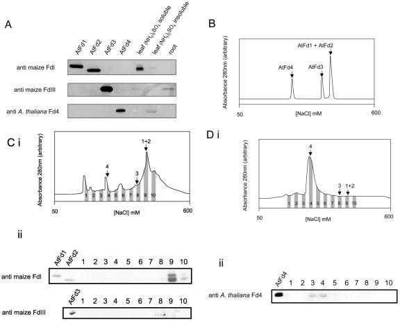
In higher plant plastids, ferredoxin (Fd) is the unique soluble electron carrier protein located in the stroma. Consequently, a wide variety of essential metabolic and signaling processes depend upon reduction by Fd. The currently available plant genomes of Arabidopsis and rice (Oryza sativa) contain several genes encoding putative Fds, although little is known about the proteins themselves. To establish whether this variety represents redundancy or specialized function, we have recombinantly expressed and purified the four conventional [2Fe-2S] Fd proteins encoded in the Arabidopsis genome and analyzed their physical and functional properties. Two proteins are leaf type Fds, having relatively low redox potentials and supporting a higher photosynthetic activity. One protein is a root type Fd, being more efficiently reduced under nonphotosynthetic conditions and supporting a higher activity of sulfite reduction. A further Fd has a remarkably positive redox potential and so, although redox active, is limited in redox partners to which it can donate electrons. Immunological analysis indicates that all four proteins are expressed in mature leaves. This holistic view demonstrates how varied and essential soluble electron transfer functions in higher plants are fulfilled through a diversity of Fd proteins.


Figures

Figure 1.
Figure 1.
Comparison of AtFd amino acid sequences with characterized Fd proteins. A, Protein sequences typical of leaf, root, and cyanobacterial [2Fe-2S] Fds aligned with Arabidopsis Fds in ClustalW 1.8 (http://searchlauncher.bcm.tmc.edu/multi-align/multialign.html) and compared in BOXSHADE 3.21 (http://www.ch.embnet.org/software/BOX-_form.html). Transit peptides are shown in lowercase and mature proteins in capitals. All transit peptide cleavage sites are as resolved through chemical sequencing of mature N termini (spinach [Spinacia oleracea] and radish [Raphanus sativus] available on http://www.ncbi.nlm.nih.gov; see Hase et al. [1991a] for maize sequences), except those of Arabidopsis, rice, and sweet orange, which were estimated by comparison with known mature proteins. White on black residues are those conserved in more than nine of the sequences analyzed. B, Fd phylogenetic tree. An alignment of mature Fd amino acid sequences, lacking transit peptides, was generated in ClustalW 1.8 as for A. This alignment is presented as a Phylogram.
Figure 2.
Figure 2.
Physical properties of AtFd proteins. A, Separation of purified mature recombinant proteins. Left panel, SDS-PAGE (1 μg protein lane-1); right panel, native gradient PAGE (20 μg lane-1). Lanes were 1, AtFd1; 2, AtFd2; 3, AtFd3; and 4, AtFd4. B, Absorption spectra: AtFd1, bold gray; AtFd2, bold black; AtFd3, light gray; and AtFd4, dark gray.
Figure 3.
Figure 3.
Redox potential. A, Cyclic voltammograms taken in 100 μm solutions of AtFd1 (bold gray), AtFd2 (bold black), AtFd3 (fine gray), and AtFd4 (fine black), versus a standard hydrogen electrode. Results are typical of three independent measurements. B, Comparison of calculated potentials with known measurements of NADPH and maize proteins: FdI, Matsumura et al. (1999); FdIII, Akashi et al. (1999); leaf FNR (L-FNR) and root FNR (R-FNR), Aliverti et al. (2001). sd was less than 5 mV in all cases.
Figure 4.
Figure 4.
Electron transfer activity. A range of concentrations of AtFd1 (black circles), AtFd2 (white circles), AtFd3 (black squares), AtFd4 (white squares) were used to mediate electron flow: A, from photoreduced PSI to leaf FNR (L-FNR); B, from NADPH reduced L-FNR to cytochrome c; C, from NADPH reduced root FNR (R-FNR) to cytochrome c; D, from NADPH reduced R-FNR to SiR. Results are single data sets representative of three to four independent measurements.
Figure 5.
Figure 5.
Detection of AtFd proteins in planta. A, Western blots using antibodies specifically reacting with leaf type Fds (raised against maize FdI), root type Fds (raised against maize FdIII), and AtFd4 (raised against AtFd4). Control lanes were loaded with 200 nmol of recombinant proteins, except for detection by anti-AtFd4, where 20 nmol of AtFd4 was used. Mature Arabidopsis leaf proteins were separated into fractions soluble and insoluble in 70% saturated (NH4)2SO4, and gels were loaded with the equivalent to 12 μg of total leaf protein, except when detecting with anti-AtFd4, where 90 μg was used. Twenty micrograms of Arabidopsis root protein was loaded. B, Elution profiles of recombinant AtFd1, AtFd2, AtFd3, and AtFd4 from an anion exchange column, over a salt gradient (based on conductivity of the eluent). C, i, Elution profile of Arabidopsis leaf proteins soluble in 70% saturated (NH4)2SO4. Arrows indicate relative elution positions of recombinant proteins: 1 + 2, AtFd1 and AtFd2; 3, AtFd3; 4, AtFD4. ii, Western blots to detect putative AtFd1 and AtFd2 after SDS-PAGE (top) and putative AtFd3 after gradient native PAGE (bottom). Two hundred nanograms of recombinant proteins was used as controls. D, i, Elution of Arabidopsis leaf proteins insoluble in 70% saturated (NH4)2SO4. Arrows as described above. ii, Labeled fractions were separated by SDS-PAGE and challenged with anti-AtFd in the western blot displayed. Twenty nanograms of recombinant AtFd4 was used as a control. All western blots are representative of two to three separate experiments.
Figure 6.
Figure 6.
Design of oligonucleotides for Fd gene synthesis. Sequences designed for the terminal coding and template oligonucleotide (oligo) pair at the 5′ end of the AtFd2 gene are shown. White on black nucleotides are those altered from genomic sequence. Full sequences of all oligos are given in supplementary information.

References

    1. Akashi T, Matsumura T, Ideguchi T, Iwakiri K, Kawakatsu T, Taniguchi I, Hase T (1999) Comparison of the electrostatic binding sites on the surface of ferredoxin for two ferredoxin-dependent enzymes, ferredoxin-NADP(+) reductase and sulfite reductase. J Biol Chem 274: 29399-29405 - PubMed
    1. Aliverti A, Faber R, Finnerty CM, Ferioli C, Pandini V, Negri A, Karplus PA, Zanetti G (2001) Biochemical and crystallographic characterization of ferredoxin-NADP(+) reductase from nonphotosynthetic tissues. Biochemistry 40: 14501-14508 - PubMed
    1. Aliverti A, Hagen WR, Zanetti G (1995) Direct electrochemistry and EPR spectroscopy of spinach ferredoxin mutants with modified electron transfer properties. FEBS Lett 368: 220-224 - PubMed
    1. Aoki K, Yamamoto M, Wada K (1998) Photosynthetic and heterotrophic ferredoxin isoproteins are colocalized in fruit plastids of tomato. Plant Physiol 118: 439-449 - PMC - PubMed
    1. Arabidopsis Genome Initiative (2000) Analysis of the genome sequence of the flowering plant Arabidopsis thaliana. Nature 408: 796-815 - PubMed

Publication types

MeSH terms