. 2003 Dec 17;23(37):11759-69.
doi: 10.1523/JNEUROSCI.23-37-11759.2003.

Activity-dependent redistribution and essential role of cortactin in dendritic spine morphogenesis

Affiliations

Activity-dependent redistribution and essential role of cortactin in dendritic spine morphogenesis

Heike Hering et al. J Neurosci. .

Abstract

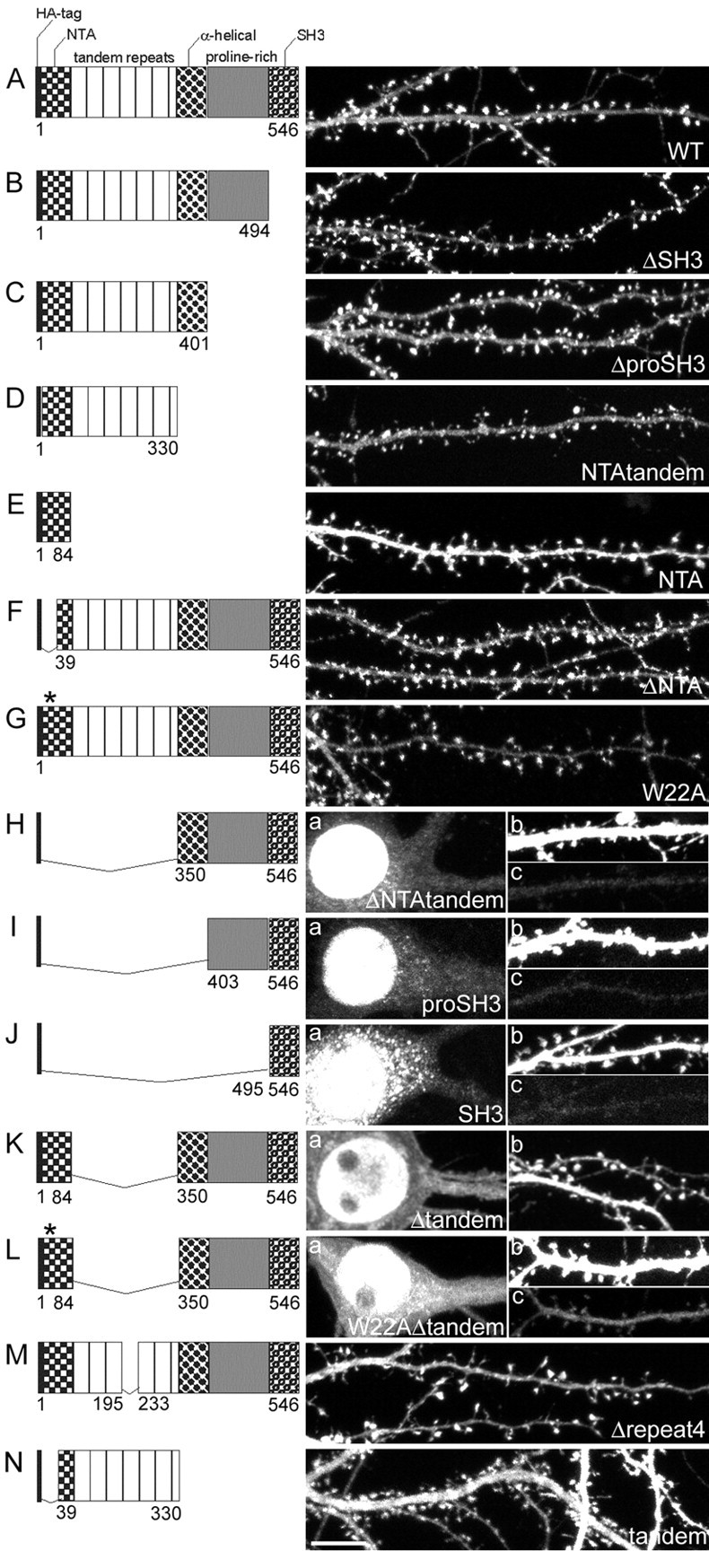
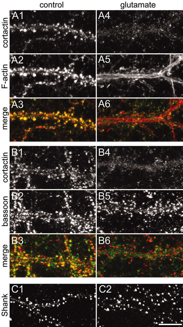
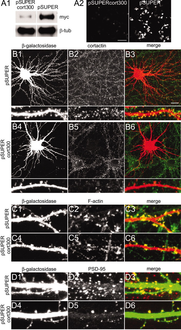
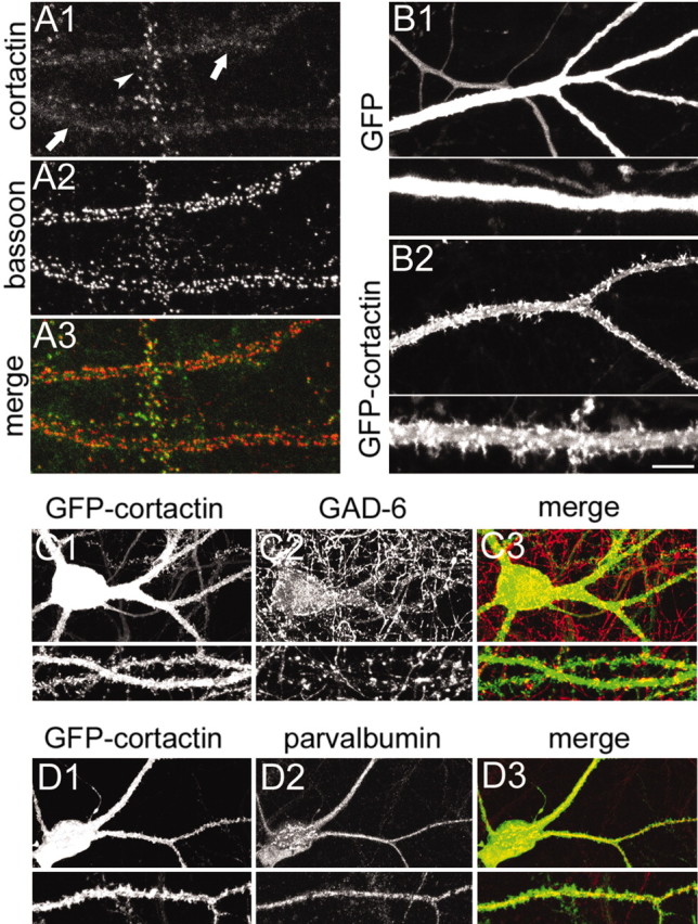
The number and shape of dendritic spines are influenced by activity and regulated by molecules that organize the actin cytoskeleton of spines. Cortactin is an F-actin binding protein and activator of the Arp2/3 actin nucleation machinery that also interacts with the postsynaptic density (PSD) protein Shank. Cortactin is concentrated in dendritic spines of cultured hippocampal neurons, and the N-terminal half of the protein containing the Arp2/3 and F-actin binding domains is necessary and sufficient for spine targeting. Knockdown of cortactin protein by short-interfering RNA (siRNA) results in depletion of dendritic spines in hippocampal neurons, whereas overexpression of cortactin causes elongation of spines. In response to synaptic stimulation and NMDA receptor activation, cortactin redistributes rapidly from spines to dendritic shaft, correlating with remodeling of the actin cytoskeleton, implicating cortactin in the activity-dependent regulation of spine morphogenesis.


Figures

Figure 2.
Figure 2.
Domain requirements for spine targeting of cortactin. Micrographs show the distribution of transfected wild-type and mutant cortactin constructs (all HA-tagged) in hippocampal neurons (DIV20; 10 d after transfection). The domain structure of the transfected cortactin mutants is shown to the left of each image. The numbers indicate the first and last amino acid for each construct. An asterisk indicates the W22A point mutation. For neurons cotransfected with β-galactosidase and HA-tagged WT (A), ΔSH3 (B), ΔproSH3 (C), NTAtandem (D), ΔNTA (F), W22A (G), Δrepeat4 (M), or tandem (N), only the distribution of cortactin construct (HA staining) is shown. For neurons cotransfected with β-galactosidase and HA-tagged ΔNTAtandem (H), proSH3 (I), SH3 (J), Δtandem (K), and W22AΔtandem (L), the HA staining pattern is shown for the soma (a in H-L) and the dendrites (b in K and c in H-J, L). The outline of the same transfected dendrite (β-galactosidase staining) is shown in b in H-J and L. Scale bar, 10 μm.
Figure 5.
Figure 5.
Glutamate-induced redistribution of cortactin from dendritic spines. All images were taken from hippocampal neurons at DIV20. A, Dendrites of a spiny neuron were double-stained for cortactin with antibody H-191 (A1, A4) and F-actin (A2, A5) in the absence of stimulation (A1-A3) or after stimulation with 100 μm glutamate for 15 min (A4-A6). Color merges are shown in A3 and A6. B, Dendrites of a spiny neuron were double-stained for cortactin with antibody H-191 (B1, B4) and bassoon (B2, B5) in the absence or presence of 100 μm glutamate for 15 min. Color merges are shown in B3 and B6. C, Dendrites of a spiny neuron were stained for Shank in the absence (C1) or presence (C2) of 100 μm glutamate for 15 min. Scale bar, 10 μm.
Figure 3.
Figure 3.
Changes in spine morphology induced by cortactin mutants. All images were taken from hippocampal neurons at DIV20 after 10 d of transfection with β-galactosidase (A), HA-tagged WT (B), ΔSH3 (C), ΔproSH3 (D), or NTAtandem (E). The cumulative distribution of spine width (F) and length (G) in neurons transfected with the constructs indicated in A-E is shown. The key for the graph in F also applies to G. H, Dendrites of neurons overexpressing wild-type cortactin (WT; green) or β-galactosidase (β-gal; green) costained for Shank (red). Scale bar (in E): A-E, H, 10 μm.
Figure 1.
Figure 1.
siRNA inhibition of cortactin causes loss of spines. A1, COS-7 cells transfected with myc-tagged murine cortactin plus pSUPERcort300 or pSUPER vector control were immunoblotted for myc and β-tubulin, as indicated. A2, GFP fluorescence in COS-7 cells cotransfected with GFP-tagged murine cortactin plus pSUPERcort300 or pSUPER. B-D, Hippocampal neurons at DIV16 cotransfected with β-galactosidase plus pSUPERcort300 or pSUPER were double-stained as indicated for cortactin (antibody H-191; B2, B5) and β-galactosidase (B1, B4); β-galactosidase (C1, C4) and rhodamine-phalloidin (C2, C5); β-galactosidase (D1, D4) and PSD-95 (D2, D5). Color merges of the double-labeled images are shown in B3, C3, D3 and B6, C6, D6 for each condition. Scale bars: A2, 200 μm; (in B3) B1-B6, 20 μm; for high-magnification micrographs in B1-B6, C, D, 5 μm.
Figure 4.
Figure 4.
Effects of cortactin overexpression in aspiny interneurons. All images were taken from neurons at DIV20. A, Dendrites of an aspiny interneuron (arrow) and a spiny pyramidal neuron (arrowhead) costained for cortactin with antibody H-191 (A1) and bassoon (A2). A3, The color merge of the two images. B, Dendrites of interneurons infected with Sindbis virus expressing GFP (B1) or GFP-cortactin (B2) for 16-20 hr. Bottom panels show dendrites at higher magnification. C, Hippocampal neuron infected with Sindbis virus expressing GFP-cortactin (C1) and costained for GAD (C2), merged in C3. D, Hippocampal neuron infected with Sindbis virus expressing GFP-cortactin (D1) and costained for parvalbumin (D2), merged in D3. Scale bar (in B2): C1-C3, D1-D3, 20 μm; A1-A3, B1, B2, and high-magnification views of C, D, 10 μm; high-magnification views of B, 5 μm.
Figure 6.
Figure 6.
Dynamics of cortactin in dendritic spines. Shown are time-lapse image series for GFP-cortactin or GFP-Shank in transfected neurons. Pixels saturated by high GFP fluorescence intensity are shown in red; pixels without fluorescence signal are shown in blue. Fluorescence intensities below saturation are shown in gray scale. Images were taken every 3 min for 1 hr (only every second image is shown; time stamps in minutes). Asterisks indicate the application of 100 μm glutamate ∼100 sec before the image acquisition. A, Dendrite of a spiny neuron (DIV20) 16-20 hr after infection with GFP-cortactin Sindbis virus. Arrows point to protrusions emerging from spine heads. B, Dendrites of a spiny neuron (DIV17) transfected with GFP-tagged Shank (GFP at N terminus of Shank). Scale bar, 10 μm.
Figure 7.
Figure 7.
NMDA receptor-dependent translocation of cortactin. All images were taken from hippocampal neurons at DIV20. A-E, Dendrites of spiny neurons double-stained for cortactin with antibody H-191 (A1-E1) and F-actin (A2-E2) after the application of 50 μm NMDA, 30 μm CNQX, and 1 μm TTX (A); 100 μm AMPA, 100 μm APV, and 1 μm TTX (B); 500 μm APV and 100 μm glutamate for 15 min (C); 50 μm bicuculline and 500 μm 4-AP for 1 hr (D); or 30 μm glutamate for 15 min (E). A3-E3, Color merges of double-labeled images. F, Time-lapse series of a dendrite of a spiny neuron infected with Sindbis virus expressing GFP-cortactin. Fluorescence intensity is represented as in Figure 6; time stamps are in minutes. Asterisks indicate the application of bicuculline and 4-AP ∼100 sec before the image was taken. G, Dendrites of spiny neurons stained with the cortactin antibody H-191 in the presence of 2 μm jasplakinolide (G1) or 2 μm jasplakinolide and 100 μm glutamate (G2). H, Dendrites of hippocampal neurons double-stained for cortactin with antibody H-191 and MAP2 after the application of 30 μm glutamate for 15 min (H1), 50 μm bicuculline and 500 μm 4-AP for 4 hr (H2), and control (H3). Scale bar (in D): A-E, G, 10 μm; F, 5 μm; H, 20 μm.

References

    1. Bailly M, Ichetovkin I, Grant W, Zebda N, Machesky LM, Segall JE, Condeelis J ( 2001) The F-actin side binding activity of the Arp2/3 complex is essential for actin nucleation and lamellipod extension. Curr Biol 11: 620-625. - PubMed
    1. Benson DL, Watkins FH, Steward O, Banker G ( 1994) Characterization of GABAergic neurons in hippocampal cell cultures. J Neurocytol 23: 279-295. - PubMed
    1. Bindokas VP, Miller RJ ( 1995) Excitotoxic degeneration is initiated at non-random sites in cultured rat cerebellar neurons. J Neurosci 15: 6999-7011. - PMC - PubMed
    1. Birukov KG, Birukova AA, Dudek SM, Verin AD, Crow MT, Zhan X, DePaola N, Garcia JG ( 2002) Shear stress-mediated cytoskeletal remodeling and cortactin translocation in pulmonary endothelial cells. Am J Respir Cell Mol Biol 26: 453-464. - PubMed
    1. Brewer GJ, Torricelli JR, Evege EK, Price PJ ( 1993) Optimized survival of hippocampal neurons in B27-supplemented Neurobasal, a new serum-free medium combination. J Neurosci Res 35: 567-576. - PubMed

Publication types

LinkOut - more resources