. 2004 Jan;239(1):118-26.
doi: 10.1097/01.sla.0000103067.10695.74.

Venous claudication in iliofemoral thrombosis: long-term effects on venous hemodynamics, clinical status, and quality of life

Affiliations

Venous claudication in iliofemoral thrombosis: long-term effects on venous hemodynamics, clinical status, and quality of life

Konstantinos T Delis et al. Ann Surg. 2004 Jan.

Abstract

Objective: We evaluated the long-term impact of iliofemoral thrombosis (I-FDVT) on walking capacity, venous hemodynamic status, CEAP class, venous clinical severity, and quality of life, and determined the prevalence of venous claudication.

Materials and methods: All patients with prior I-FDVT, assessed at our institution since 1990, were called for follow-up. Those with walking impairment due to arterial disease (ABI < 1.0 postexercise) or unrelated causes and those thrombectomized or thrombolyzed were excluded; 39 patients (22-83 years, median 46 years) were included. Median follow-up was 5 years (range 1-23 years). Investigation included classification in CEAP and Venous Clinical Severity Scoring (VCSS) systems, air-plethysmography (outflow fraction [OF], venous filling index [VFI], residual volume fraction [RVF]) and venous duplex, treadmill (3.5 km/h, 10%) to determine initial (ICD) and absolute (ACD) claudication distances, and quality of life assessment (SF-36). Nonaffected limbs of patients with unilateral I-FDVT (37 of 39) comprised the control group. Data are presented as median and interquartile range.

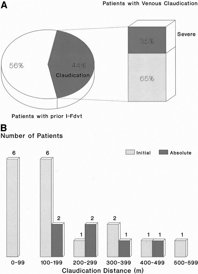
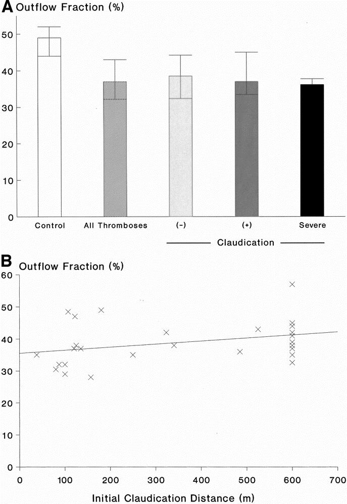
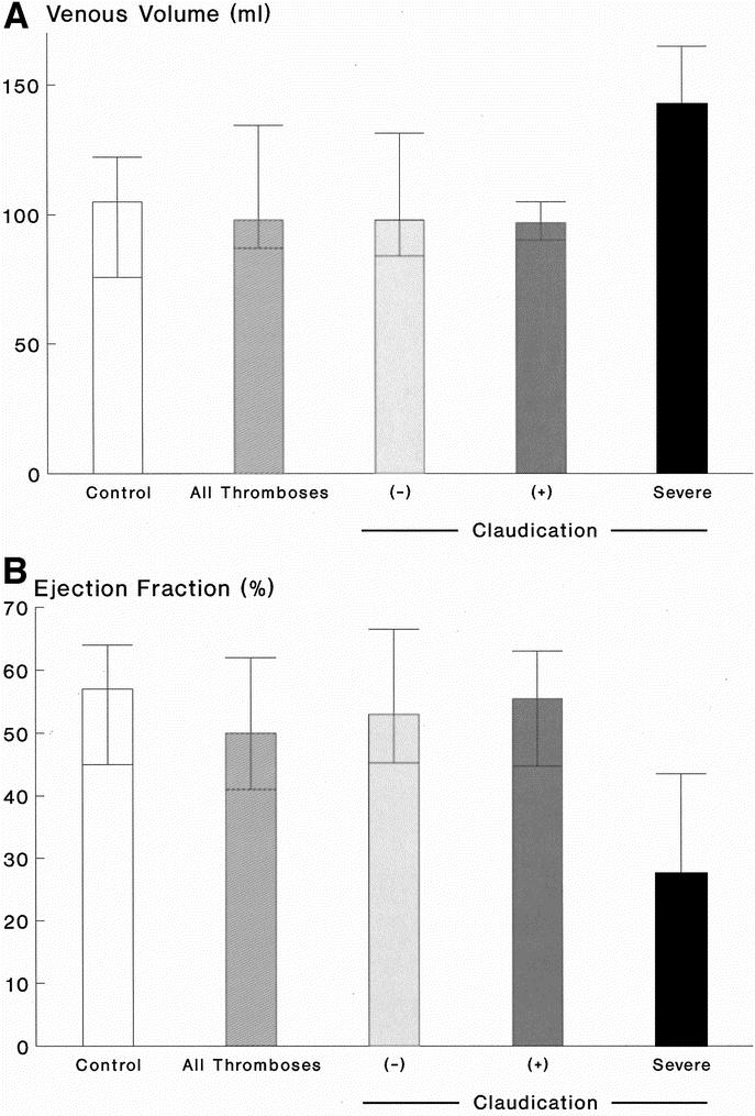
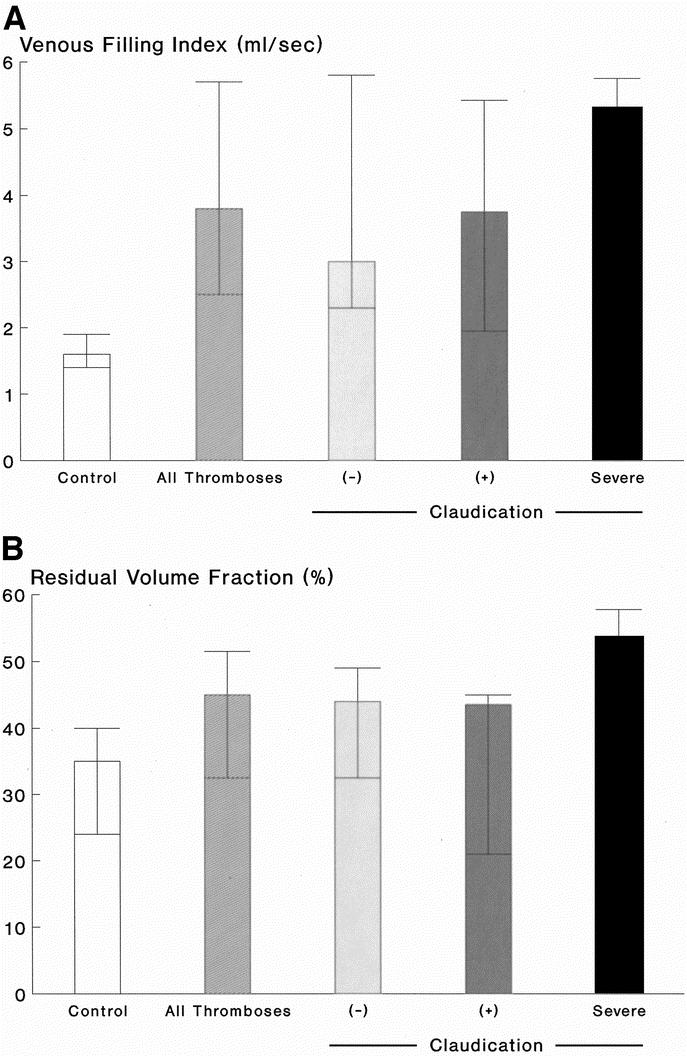
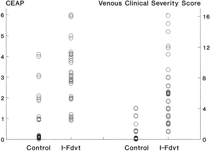
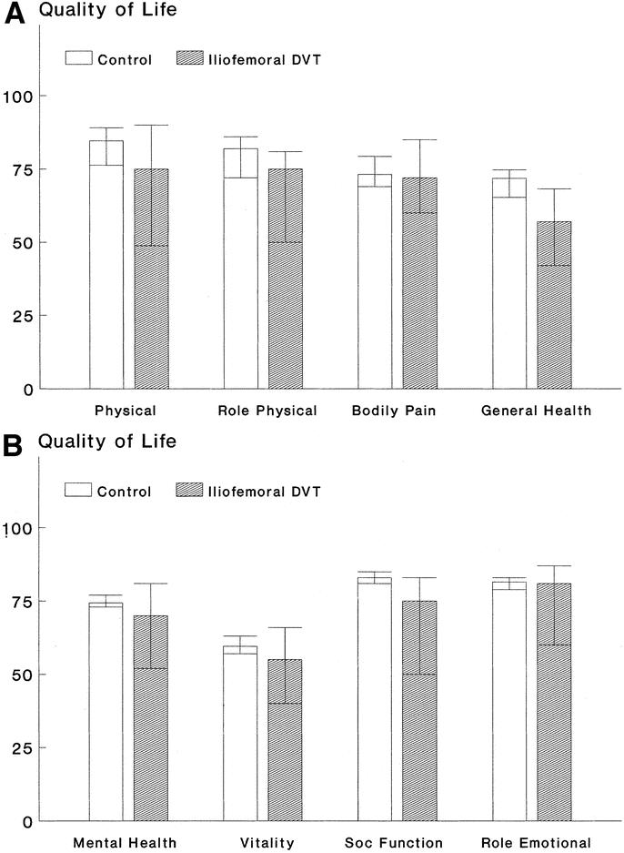
Results: A total of 81% of limbs with I-FDVT had superficial and deep reflux and 19% superficial reflux; reflux in control limbs was 29.7% (P < 0.001) and 27% (P > 0.2), respectively; 43.6% (17 of 39; 95% CI, 27-60%) of patients developed venous claudication ipsilateral to I-FDVT (ICD: 130 m, range 105-268 m), compelling 15.4% (6 of 39; 95% CI, 3.5-27%) to discontinue treadmill (ACD: 241 m, range 137-298 m). Limbs with prior I-FDVT had a lower OF (37%, range 32.2-43%; P < 0.001), abnormally higher VFI (3.8 mL/s, range 2.5-5.7 mL/s; P < 0.001), and RVF (45%, range 32.5-51.5%; P = 0.006), and clinical impairment in CEAP and VCSS systems (P < 0.0001). Patients with I-FDVT had impaired physical functioning (P = 0.02) and role (P = 0.033), general health (P = 0.001), social function (P = 0.047), and mental health (P = 0.043).

Conclusions: A total of 43.6% of those with prior I-FDVT developed venous claudication compelling interruption of walking in 15.4%. Prior I-FDVT caused outflow impairment and a large residual venous volume and reflux, resulting in marked clinical and quality of life compromise. Standardized challenge enabled discrimination of those with clinically relevant impairment.


Figures

None
FIGURE 1. A: The prevalence of intermittent venous claudication among 39 patients with prior I-FDVT, after a median 5-year (range 1–23 years) follow-up period; 43.6% (17 of 39) developed intermittent claudication at a median initial claudication distance of 130 m (interquartile range 105–268 m), the severity of which compelled 6 of them [35% (6 of 17)] or 15.4% (6 of 39) of the total to discontinue the treadmill at a median absolute claudication distance of 241 m (interquartile range 137–298 m). B: Distribution of initial and absolute claudication distances among the 17 patients of our study population with prior iliofemoral thrombosis and intermittent venous claudication.
None
FIGURE 2. A: Outflow fraction [OF] (%) in 37 control limbs and 41 limbs with prior I-FDVT (P < 0.001), on the left. On the right, the limbs with prior I-FDVT are subdivided into those with (+) and without (–) claudication, and those with severe claudication compelling cessation of walking; differences among the three subgroups were not significant (P = 0.58). Data are expressed as median and interquartile ranges. B: Outflow fraction (%) in limbs with prior I-FDVT plotted against the initial claudication distance (m) (r = 0.43, P = 0.018, Spearman’s two-tailed). Patients with shortness of breath were excluded.
None
FIGURE 3. A: Venous volume [VV] (mL) in 37 control limbs and 41 limbs with prior I-FDVT (P = 0.27), on the left. On the right, the limbs with prior I-FDVT are subdivided into those with (+) and without (–) claudication, and those with severe claudication compelling cessation of walking; differences among the three subgroups were not significant (P = 0.58). Data are expressed as median and interquartile ranges. B: Ejection fraction [EF] (%) in 37 control limbs and 41 limbs with prior I-FDVT (P = 0.13), on the left. On the right, the limbs with I-FDVT are subdivided into those with (+) and without (–) claudication, and those with severe claudication compelling cessation of walking; differences among the three subgroups were significant overall (P = 0.014) [(–) vs. severe, P = 0.003; (+) vs. severe, P = 0.016]. Data are expressed as median and interquartile ranges.
None
FIGURE 4. A: Venous filling index [VFI] (mL/s) in 37 control limbs and 41 limbs with prior I-FDVT (P < 0.001), on the left. On the right, the limbs with prior I-FDVT are subdivided into those with (+) and without (–) claudication, and those with severe claudication compelling cessation of walking; differences among the three subgroups were not significant overall (P = 0.22) [(–) vs. severe, P = 0.09; (+) vs. severe, P = 0.18]. Data are expressed as median and interquartile ranges. B: Residual volume fraction [RVF] (%) in 37 control limbs and 41 limbs with prior I-FDVT (P = 0.006), on the left. On the right, the limbs with prior I-FDVT are subdivided into those with (+) and without (–) claudication, and those with severe claudication compelling cessation of walking; differences among the three subgroups were significant overall (P = 0.05) [(–) vs. severe, P = 0.03; (+) vs. severe, P = 0.03]. Data are expressed as median and interquartile ranges.
None
FIGURE 5. Clinical stratification of 41 limbs with prior I-FDVT and 37 control limbs according to the CEAP and the VCSS (venous clinical severity scoring) systems. Limbs with prior I-FDVT were significantly worse than the control limbs in both systems (both P < 0.0001).
None
FIGURE 6. Quality of life assessment of 39 patients with prior I-FDVT according to the Short-Form 36 health survey questionnaire, in comparison with an equal number of healthy individuals matched for age and sex. Patients with prior I-FDVT had a significantly worse perception of their physical function (P = 0.02), physical role (P = 0.033), general health (P = 0.001) [a], social function (P = 0.047), and mental health (P = 0.043) [b]; there was no difference in the bodily pain (P = 0.57) [a], vitality (P = 0.085), and role emotional (P = 0.4).

References

    1. Gjores JE. The incidence of venous thrombosis and its sequelae in certain districts of Sweden. Acta Chir Scand. 1956;(suppl 206). - PubMed
    1. Mavor GE, Galloway JM. Iliofemoral venous thrombosis: pathological considerations and surgical management. Br J Surg. 1969;56:45–59. - PubMed
    1. Prandoni P, Lensing AW, Cogo A, et al. The long-term clinical course of acute deep venous thrombosis. Ann Intern Med. 19961;125:1–7. - PubMed
    1. Plate G, Eklof B, Norgren L, et al. Venous thrombectomy for iliofemoral vein thrombosis: 10-year results of a prospective randomised study. Eur J Vasc Endovasc Surg. 1997;14:367–374. - PubMed
    1. Kahn SR, Solymoss S, Lamping DL, et al. Long-term outcomes after deep vein thrombosis: postphlebitic syndrome and quality of life. J Gen Intern Med. 2000;15:425–429. - PMC - PubMed

MeSH terms