. 2004 Jan 14;23(1):160-8.
doi: 10.1038/sj.emboj.7600016. Epub 2003 Dec 11.

Identification of a redox-regulated chaperone network

Affiliations

Identification of a redox-regulated chaperone network

Jörg H Hoffmann et al. EMBO J. .

Abstract

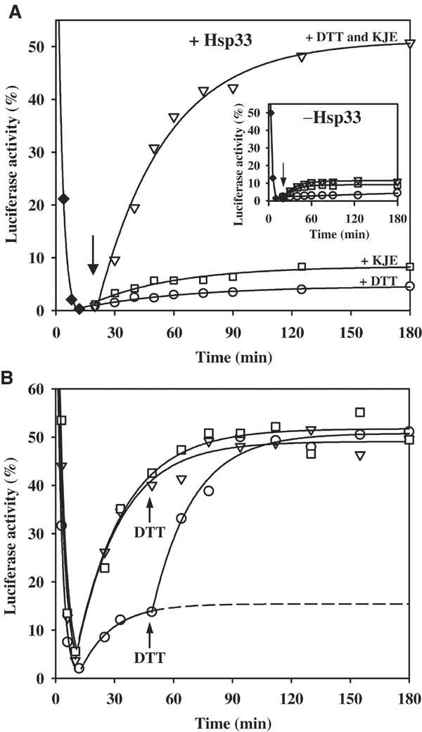
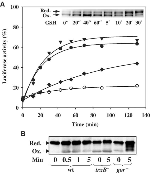
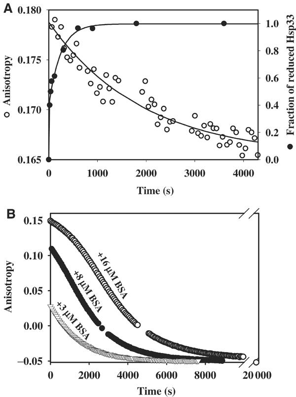
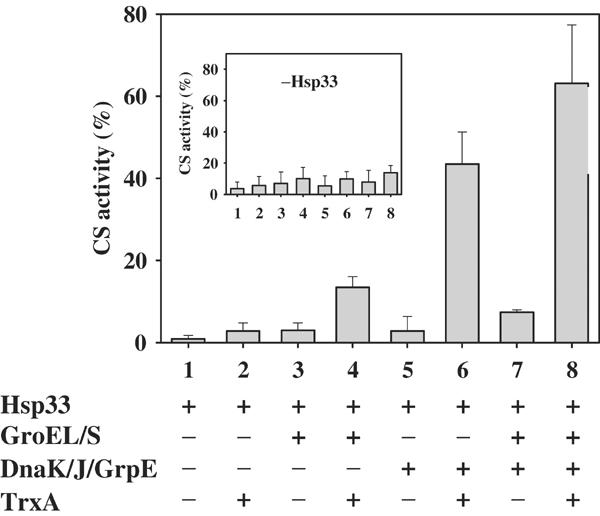
We have identified and reconstituted a multicomponent redox-chaperone network that appears to be designed to protect proteins against stress-induced unfolding and to refold proteins when conditions return to normal. The central player is Hsp33, a redox-regulated molecular chaperone. Hsp33, which is activated by disulfide bond formation and subsequent dimerization, works as an efficient chaperone holdase that binds to unfolding protein intermediates and maintains them in a folding competent conformation. Reduction of Hsp33 is catalyzed by the glutaredoxin and thioredoxin systems in vivo, and leads to the formation of highly active, reduced Hsp33 dimers. Reduction of Hsp33 is necessary but not sufficient for substrate protein release. Substrate dissociation from Hsp33 is linked to the presence of the DnaK/DnaJ/GrpE foldase system, which alone, or in concert with the GroEL/GroES system, then supports the refolding of the substrate proteins. Upon substrate release, reduced Hsp33 dimers dissociate into inactive monomers. This regulated substrate transfer ultimately links substrate release and Hsp33 inactivation to the presence of available DnaK/DnaJ/GrpE, and, therefore, to the return of cells to non-stress conditions.


Figures

Figure 1
Figure 1
Reduction of Hsp33 is not sufficient to cause substrate release from Hsp33. Light scattering measurements of luciferase (140 nM) were performed either in the absence (a) or presence of 210 nM active Hsp33 dimers (b, c) at 43°C. At the time point indicated by the arrow, 5 mM DTT was added to reactions a and b, and the incubation was continued at 43°C. Inset: DTT induces the fast reduction of Hsp33's thiol groups. Dimeric oxidized Hsp33 in complex with thermally unfolded luciferase (left panel) or oxidized Hsp33 alone (right panel) was incubated with 5 mM DTT at 25°C. At the time points indicated, aliquots were withdrawn and free thiol groups were covalently modified with the 500 Da molecule AMS as described. Similar results were obtained at 43°C, albeit with significantly faster reduction rates.
Figure 2
Figure 2
Substrate shuttling from the Hsp33 holdase to the DnaK/DnaJ/GrpE foldase. (A) Hsp33 transfers substrates to the DnaK/DnaJ/GrpE system for refolding. Luciferase (80 nM) was thermally unfolded in the presence of a five-fold molar excess of active Hsp33 dimers for 10 min at 43°C (⧫). Then, the temperature was shifted to 25°C. After 10 min of incubation at 25°C (as indicated by the arrow), either (○) 5 mM DTT, (□) DnaK/DnaJ/GrpE in a 5:2:5 molar ratio of the monomers or (▿) a combination of 5 mM DTT and DnaK/DnaJ/GrpE (5:2:5) was added to the reaction. The reactivation reaction of luciferase was monitored following luminescence. Inset: Substrate transfer and luciferase refolding requires the presence of active Hsp33 during inactivation. Luciferase (80 nM) was thermally unfolded for 10 min at 43°C without any chaperones (⧫). At 10 min after shift of the temperature to 25°C, either (○) 5 mM DTT, (□) DnaK/ DnaJ/GrpE in a 5:2:5 molar ratio of the monomers or a combination of (▿) 5 mM DTT and DnaK/DnaJ/GrpE (5:2:5) was added to the reaction and the reactivation reaction was monitored. (B) Reduction of Hsp33 and substrate transfer to the DnaK/DnaJ/GrpE system is not rate limiting for luciferase refolding. Luciferase (80 nM) was thermally unfolded (43°C) with DnaK/DnaJ/GrpE (molar ratio 5:2:5) either in the (□) presence of 5 mM DTT and a 10-fold molar excess of reduced, monomeric Hsp33, or in the (▿) absence of Hsp33 or (○) in the presence of a five-fold molar excess of active Hsp33 dimers. After 10 min at 43°C, the temperature was shifted to 25°C. At the time point indicated, 5 mM DTT was added to the reactions that did not contain DTT.
Figure 3
Figure 3
Identification of the physiological reductants of Hsp33. (A) Reduction of Hsp33 is catalyzed by thioredoxin in a TrxB- and NADPH-dependent manner in vitro. Luciferase (140 nM) was thermally unfolded in the presence of DnaK/DnaJ/GrpE (molar ratios 5:2:5) and a five-fold molar excess of active Hsp33 dimer for 8 min at 43°C. After 12 min of incubation at room temperature, the reaction was shifted to 30°C (t=0 min) and the incubation reaction was continued in the (○) absence of any additives or in the presence of either (▾) 5 mM DTT, (⧫) 5 mM GSH or (•) 2.8 μM TrxA, 70 nM TrxB, 50 μM NADPH. Only the recovery period at 30°C is shown. Inset: Physiological concentrations of reduced glutathione are not capable of quickly reducing oxidized Hsp33 dimers. Oxidized Hsp33 dimers (3 μM) were incubated in the presence of 5 mM GSH at 30°C. At the time points indicated, aliquots were removed and thiol trapping with AMS was performed as described. (B) Glutaredoxin and thioredoxin systems reduce Hsp33 in vivo. DHB4 wild-type, WM93 (ΔtrxB) or WP840 (Δgor) cells were grown at 30°C until an OD600 of 0.5 was reached. Then, the cells were shifted to 45°C. At 1 min after the shift, a sample was taken (t=0) and H2O2 was added (4 mM). Further samples were taken at the indicated time points. In vivo thiol trapping with AMS was performed and the samples were processed as described.
Figure 4
Figure 4
Identification of a stable Hsp33 intermediate: the reduced, active Hsp33 dimer. (A) Reduced Hsp33 dimers are kinetically stable. Oxidized Hsp33 dimers (3 μM) were incubated in 40 mM HEPES-KOH, 20 mM KCl, pH 8.0, at 20°C. A stable anisotropy signal at λex=280 nm and λem=350 nm was detected. To monitor (○) dissociation of reduced Hsp33 dimers, 5 mM DTT was added to the reaction. The anisotropy signal decreased and approached the lower signal of monomeric, reduced Hsp33. The curve was fitted according to a pseudo-first-order reaction and a rate constant of 5.2 × 10⁻⁴ s−1 was determined. A very similar rate constant was obtained when 15 μM Hsp33 dimers were used instead (4.7 × 10⁻⁴ s−1). To monitor the (•) reduction of Hsp33's thiol groups under these conditions, aliquots were taken and AMS trapped as described. The proteins were separated on 14% SDS–PAGE and the fraction of reduced Hsp33 protein was determined using a densitometer. A rate constant of 0.018 s−1 was determined for the reduction process of Hsp33. (B) Substrate proteins stabilize reduced Hsp33 dimers and delay inactivation of Hsp33. Oxidized Hsp33 dimers (3 μM) labeled with the fluorescent dye Oregon-green were incubated in 40 mM HEPES-KOH, 20 mM KCl, pH 8.0, at 20°C in the presence of (▿) 3 μM BSA, (•) 8 μM BSA or (○) 16 μM BSA. To reduce the dimeric Hsp33–BSA complexes, 5 mM DTT was added to the reaction, and the change in anisotropy signal was followed at λex=506 nm and λem=524 nm. Thiol trapping experiments showed that the rate of Hsp33 reduction was not significantly influenced by the presence of 16 μM BSA. Analysis of the apparent dissociation rate of fluorescently labeled Hsp33 dimers in the absence of BSA revealed a rate constant that was very similar to unlabeled Hsp33 (5.6 × 10⁻⁴ s−1).
Figure 5
Figure 5
In vitro reconstitution of a redox-regulated multichaperone network. Chemically denatured CS was diluted to a final concentration of 75 nM into renaturation buffer (40 mM HEPES-KOH, pH 8.0, 10 mM KCl, 2 mM MgATP) containing a five-fold molar excess of active Hsp33 dimers. At 20 min after the start of the renaturation, various combinations of reduced TrxA (3 μM), DnaK:DnaK:GrpE (0.4 μM:0.16 μM:0.4 μM) and GroEL14:GroES7 (0.15 μM:0.5 μM) were added. The incubation reaction was continued for 160 min and the activity of CS was analyzed. The inset shows the identical experiment in the absence of dimeric Hsp33.
Figure 6
Figure 6
A redox-regulated chaperone network. Under nonstress conditions, Hsp33 is monomeric and all four cysteines are reduced and coordinate one zinc ion. Hsp33 is in its low-affinity binding state. Upon exposure to heat and oxidative stress, two intramolecular disulfide bonds form in Hsp33, the zinc is released and Hsp33 dimerizes. Hsp33 is in its high-affinity binding state. It acts as a chaperone holdase and protects proteins against irreversible aggregation processes by tightly binding to unfolding protein intermediates. The thioredoxin system quickly reduces the dimeric Hsp33–substrate protein complexes. This does not cause any substrate protein release, but rather primes Hsp33 for fast inactivation once the DnaK/DnaJ/GrpE system is available. Upon return of cells to nonstress conditions, sufficient amounts of the DnaK/DnaJ/GrpE foldase system are available to release the substrate proteins from Hsp33. These substrate proteins are then refolded by the DnaK/DnaJ/GrpE foldase system alone or together with the GroEL/GroES system.

References

    1. Aslund F, Zheng M, Beckwith J, Storz G (1999) Regulation of the OxyR transcription factor by hydrogen peroxide and the cellular thiol-disulfide status. Proc Natl Acad Sci USA 96: 6161–6165 - PMC - PubMed
    1. Barbirz S, Jakob U, Glocker MO (2000) Mass spectrometry unravels disulfide bond formation as the mechanism that activates a molecular chaperone. J Biol Chem 275: 18759–18766 - PubMed
    1. Beissinger M, Buchner J (1998) How chaperones fold proteins. Biol Chem 379: 245–259 - PubMed
    1. Buchberger A, Schroder H, Buttner M, Valencia A, Bukau B (1994) A conserved loop in the ATPase domain of the DnaK chaperone is essential for stable binding of GrpE. Nat Struct Biol 1: 95–101 - PubMed
    1. Buchner J, Schmidt M, Fuchs M, Jaenicke R, Rudolph R, Schmid FX, Kiefhaber T (1991) GroE facilitates refolding of citrate synthase by suppressing aggregation. Biochemistry 30: 1586–1591 - PubMed

Publication types

MeSH terms