

Selective induction of tumor necrosis receptor factor 6/decoy receptor 3 release by bacterial antigens in human monocytes and myeloid dendritic cells
- PMID: 14688085
- PMCID: PMC343977
- DOI: 10.1128/IAI.72.1.89-93.2004
Selective induction of tumor necrosis receptor factor 6/decoy receptor 3 release by bacterial antigens in human monocytes and myeloid dendritic cells
Abstract
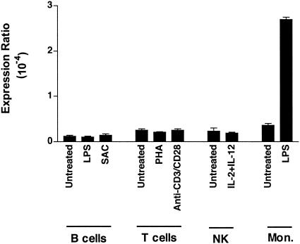
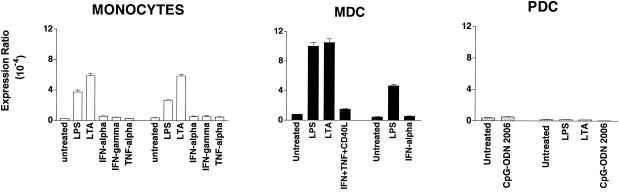
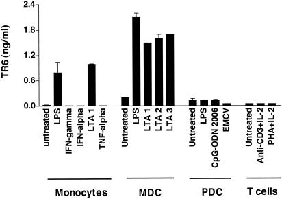
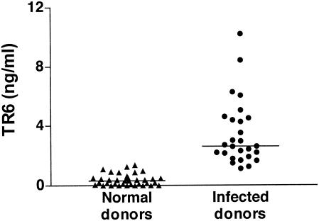
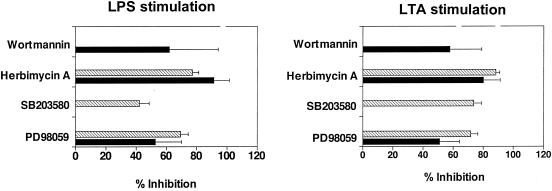
Tumor necrosis factor (TNF) receptor 6/decoy receptor 3 (TR6/DcR3) is an antiapoptosis soluble receptor of the TNF family produced by tumor cells. In this study, TR6 expression in human immune cells was investigated. TR6 mRNA and protein were detectable in selected antigen-presenting cells. Monocytes and myeloid-derived dendritic cells (MDC) released the protein exclusively following stimulation of Toll-like receptor 2 (TLR2) and TLR4 by gram-positive and gram-negative bacterial antigens. Plasmacytoid dendritic cells, activated by bacterial antigens via TLR9 or by viral infection, did not produce the protein. Similarly, activated T cells did not release TR6. The release of TR6 by MDC was dependent on the activation of p42/p44 mitogen-activated protein kinases, Src-like protein tyrosine kinases, and phosphatidylinositol 3-kinase, signaling pathways important for MDC maturation and survival. In agreement with the in vitro data, TR6 levels in serum were significantly elevated in patients with bacterial infections. Overall, these data suggest a novel role for TR6 in immune responses to bacteria.
Figures
Similar articles
-
Pattern of cytokine responses to gram-positive and gram-negative commensal bacteria is profoundly changed when monocytes differentiate into dendritic cells.Infect Immun. 2004 May;72(5):2671-8. doi: 10.1128/IAI.72.5.2671-2678.2004. Infect Immun. 2004. PMID: 15102775 Free PMC article.
-
Death decoy receptor TR6/DcR3 inhibits T cell chemotaxis in vitro and in vivo.J Immunol. 2003 Oct 1;171(7):3407-14. doi: 10.4049/jimmunol.171.7.3407. J Immunol. 2003. PMID: 14500635
-
A newly identified member of tumor necrosis factor receptor superfamily (TR6) suppresses LIGHT-mediated apoptosis.J Biol Chem. 1999 May 14;274(20):13733-6. doi: 10.1074/jbc.274.20.13733. J Biol Chem. 1999. PMID: 10318773
-
Role of MD-2 in TLR2- and TLR4-mediated recognition of Gram-negative and Gram-positive bacteria and activation of chemokine genes.J Endotoxin Res. 2000;6(5):401-5. doi: 10.1179/096805100101532243. J Endotoxin Res. 2000. PMID: 11521063 Review.
-
Toll-like receptors as adjuvant receptors.Biochim Biophys Acta. 2002 Feb 13;1589(1):1-13. doi: 10.1016/s0167-4889(01)00182-3. Biochim Biophys Acta. 2002. PMID: 11909637 Review.
Cited by
-
Programmed cell death of dendritic cells in immune regulation.Immunol Rev. 2010 Jul;236:11-27. doi: 10.1111/j.1600-065X.2010.00916.x. Immunol Rev. 2010. PMID: 20636805 Free PMC article. Review.
-
Decoy receptor 3, a novel inflammatory marker, and mortality in hemodialysis patients.Clin J Am Soc Nephrol. 2012 Aug;7(8):1257-65. doi: 10.2215/CJN.08410811. Epub 2012 May 24. Clin J Am Soc Nephrol. 2012. PMID: 22626963 Free PMC article.
-
Increased circulating levels of tumor necrosis factor-like cytokine 1A and decoy receptor 3 correlate with SYNTAX score in patients undergoing coronary surgery.J Int Med Res. 2018 Dec;46(12):5167-5175. doi: 10.1177/0300060518793787. Epub 2018 Sep 13. J Int Med Res. 2018. PMID: 30213220 Free PMC article.
-
Predictive value of decoy receptor 3 in postoperative nosocomial bacterial meningitis.Int J Mol Sci. 2014 Nov 3;15(11):19962-70. doi: 10.3390/ijms151119962. Int J Mol Sci. 2014. PMID: 25372942 Free PMC article.
-
Significance of increased expression of decoy receptor 3 in chronic liver disease.Dig Liver Dis. 2009 Aug;41(8):591-8. doi: 10.1016/j.dld.2008.11.019. Epub 2009 Feb 4. Dig Liver Dis. 2009. PMID: 19195939 Free PMC article.
References
-
- Arbibe, L., J. P. Mira, N. Teusch, L. Kline, M. Guha, N. Mackman, P. J. Godowski, R. J. Ulevitch, and U. G. Knaus. 2000. Toll-like receptor 2-mediated NF-kappa B activation requires a Rac1-dependent pathway. Nat. Immunol. 1:533-540. - PubMed
-
- Ardershna, K. M., A. R. Pizzey, S. Devereux, and A. Khwaja. 2000. The PI3 kinase, p38 SAP kinase, and NF-κB signal transduction pathways are involved in the survival and maturation of lipopolysaccharide-stimulated human monocyte-derived dendritic cells. Blood 96:1039-1046. - PubMed
-
- Bai, C., B. Connolly, M. L. Metzker, C. A. Hilliard, X. Liu, V. Sandig, A. Soderman, S. M. Galloway, Q. Liu, C. P. Austin, and C. T. Caskey. 2000. Overexpression of M68/TR6 in human gastrointestinal tract tumors independent of gene amplification and its location in a four-gene cluster. Proc. Natl. Acad. Sci. USA 97:1230-1235. - PMC - PubMed
-
- Bauer, M., V. Redecke, J. W. Ellwart, B. Ascherer, J.-P. Kremer, H. Wagner, and G. B. Lipford. 2001. Bacterial CpG-DNA triggers activation and maturation of human CD11c−, CD123+ dendritic cells. J. Immunol. 166:5000-5007. - PubMed
-
- Connolly, K., Y. H. Cho, R. Duan, J. Fikes, T. Gregorio, D. W. LaFleur, Z. Okoye, T. W. Salcedo, G. Santiago, S. Ullrich, P. Wei, K. Windle, E. Wong, X. T. Yao, Y. Q. Zhang, G. Zheng, and P. A. Moore. 2001. In vivo inhibition of Fas ligand mediated killing by TR6, a Fas ligand decoy receptor. J. Pharmacol. Exp. Ther. 298:25-33. - PubMed
Publication types
MeSH terms
Substances
LinkOut - more resources
Full Text Sources
Other Literature Sources
Medical
Miscellaneous
