. 2004 Jan;72(1):94-105.
doi: 10.1128/IAI.72.1.94-105.2004.

Role of the C-terminal lysine residues of streptococcal surface enolase in Glu- and Lys-plasminogen-binding activities of group A streptococci

Affiliations

Role of the C-terminal lysine residues of streptococcal surface enolase in Glu- and Lys-plasminogen-binding activities of group A streptococci

Anne Derbise et al. Infect Immun. 2004 Jan.

Abstract

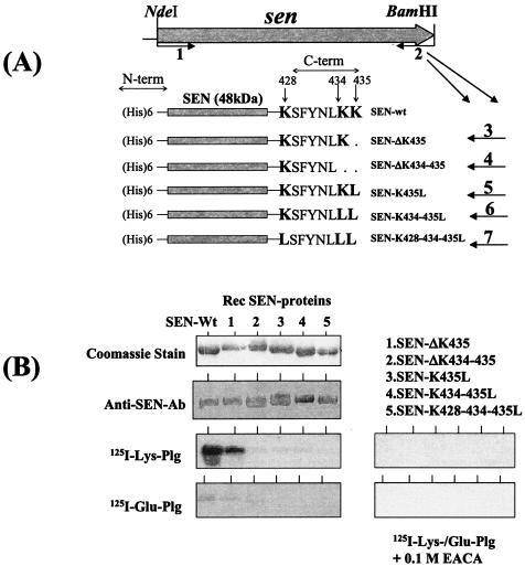
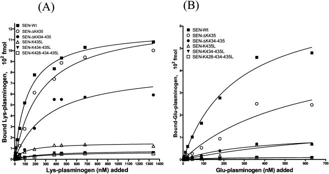
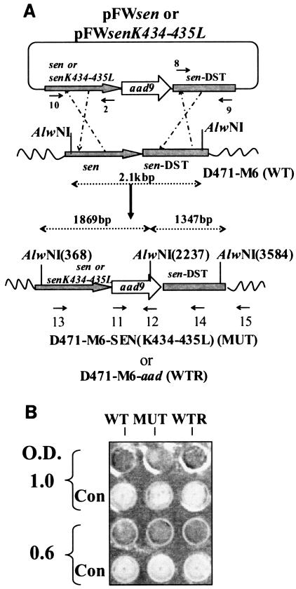
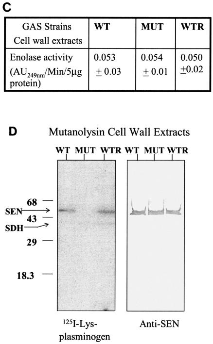
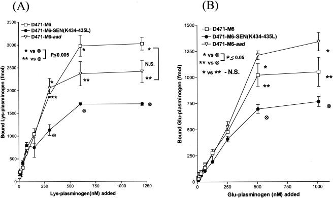
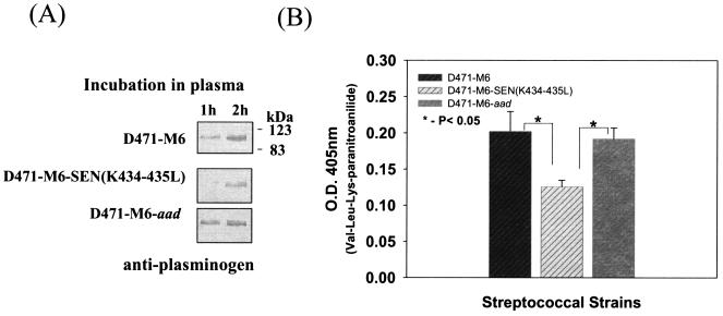
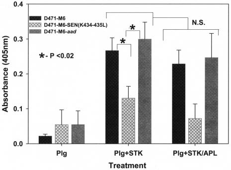
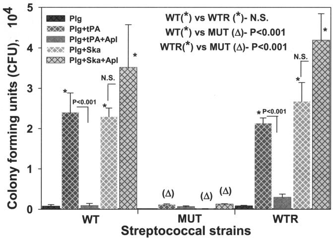
Streptococcal surface enolase (SEN) is a major plasminogen-binding protein of group A streptococci. Our earlier biochemical studies have suggested that the region responsible for this property is likely located at the C-terminal end of the SEN molecule. In the present study, the gene encoding SEN was cloned from group A streptococci M6 isolate D471. A series of mutations in the sen gene corresponding to the C-terminal region (428KSFYNLKK435) of the SEN molecule were created by either deleting one or more terminal lysine residues or replacing them with leucine. All purified recombinant SEN proteins with altered C-terminal ends were found to be enzymatically active and were analyzed for their Glu- and Lys-plasminogen-binding activities. Wild-type SEN bound to Lys-plasminogen with almost three times more affinity than to Glu-plasminogen. However, the recombinant mutant SEN proteins with a deletion of Lys434-435 or with K435L and K434-435L replacements showed a significant decrease in Glu- and Lys-plasminogen-binding activities. Accordingly, a streptococcal mutant expressing SEN-K434-435L showed a significant decrease in Glu- and Lys-plasminogen-binding activities. Biochemical and functional analyses of the isogenic mutant strain revealed a significant decrease in its abilities to cleave a chromogenic tripeptide substrate, acquire plasminogen from human plasma, and penetrate the extracellular matrix. Together, these data indicate that the last two C-terminal lysine residues of surface-exposed SEN contribute significantly to the plasminogen-binding activity of intact group A streptococci and hence to their ability to exploit host properties to their own advantage in tissue invasion.


Figures

FIG. 1.
FIG. 1.
(A) Strategy used to delete or replace the C-terminal (C-term) lysine residues (K428, K434, and K435) by site-directed mutagenesis and the resultant His-tagged recombinant SEN proteins. Primer 1 (sen-F-NdeI) is a common forward primer and contains an NdeI site. Primers 2 to 7 are reverse primers each with a restriction site for BamHI used to construct wild-type SEN (SEN-wt) and mutated SEN proteins with their C-terminal and penultimate lysine residues either deleted or replaced with leucines (see Table 1). N-term, N terminal. (B) Western blot analysis showing the antibody reactivities of different purified recombinant (Rec) SEN proteins with affinity-purified rabbit polyclonal anti-SEN antibodies (Ab) and their plasminogen (Plg)-binding activities with 125I-labeled Glu-plasminogen and 125I-labeled Lys-plasminogen in the presence or absence of 0.1 M EACA.
FIG. 2.
FIG. 2.
Solid-phase ligand-binding assay showing specific Lys-plasminogen-binding (A) and Glu-plasminogen-binding (B) activities with different His-tagged recombinant SEN proteins. The assay was performed in 96-well microtiter plates. The results were analyzed by the one-site- and two-site-binding nonlinear-curve models with GraphPad Prism version 3.03 software. The best-fit curve shown is the one-site-binding curve, as validated by the F test. The extracted affinity binding values (Kd and Bmax) for each curve are in Table 2. Each data point represents an average value of three experiments, each done with duplicate samples. SEN-Wt, wild-type SEN.
FIG. 3.
FIG. 3.
(A). Plasmid construction for replacement of the wild-type (WT) sen gene with a sen K434-435L mutation in strain D471-M6. As described in Materials and Methods, the PCR-amplified DNA fragments corresponding to the mutated sen gene and the flanking downstream regions of sen (sen DST) were digested with SalI/BamHI and PstI/NdeI, respectively, and ligated to the appropriate sites upstream and downstream of aad9 (spectinomycin resistance marker gene), respectively, into pFW5 to yield plasmid pFWsenK434-435L. The latter was then introduced into strain D471-M6 and integrated into the streptococcal chromosome via homologous recombination, resulting in the creation of a mutant (MUT) strain, D471-M6-SEN-K434-435L. A similar introduction of pFWsen created a control strain (D471-M6-aad [WTR]) that contained the aad9 gene between the wild-type sen gene and the sen DST region. Primers 10 and 15, primers 11 and 12 (derived from the aad9 gene), primers 13 and 12, and primers 11 and 15 were used to verify the chromosomal insertion of aad9 downstream of sen. The sen K435-434L mutation was verified by DNA sequence analyses of the PCR fragment obtained with primers 13 and 12. (B) Surface expression of SEN on the D471-M6, D471-M6-SEN-K434-435L, and D471-M6-aad strains. Surface expression of SEN was measured by colony blot analysis with a MultiScreen 96-well filter plate (0.22-μm-pore-size low-protein-binding Durapore membrane; Millipore) and a 96-well plate vacuum manifold. A 100-μl volume of different growth phase cultures (mid log [optical density at 600 nm = 0.6] and late log [optical density at 600 nm = 1.0]), normalized at an optical density (O.D.) at 600 nm of 1.0, were dispensed into the wells and blotted. They were then stained with anti-SEN polyclonal antibody and corresponding alkaline phosphatase conjugate and developed as shown. The control (Con) represents bacteria stained with only conjugate antibody. (C) Enolase activity of mutanolysin extracts (5 μg) of the D471-M6, D471-M6-SEN-K434-435L, and D471-M6-aad GAS strains (adjusted to an optical density at 600 nm of 1). Enolase activity was measured as described in Materials and Methods. The values shown are averages of three different experiments ± the standard deviation. (D) Plasminogen-binding activities of SEN found in the mutanolysin-extracted cell walls of streptococcal strains D471-M6, D471-M6-SEN-K434-435L, and D471-M6-aad. The reactivity of these bands with rabbit anti-SEN affinity-purified antibody is shown. SDH, streptococcal surface dehydrogenase; AU249, units of absorbance at 249 nm. The values on the left are molecular sizes in kilodaltons.
FIG. 3.
FIG. 3.
(A). Plasmid construction for replacement of the wild-type (WT) sen gene with a sen K434-435L mutation in strain D471-M6. As described in Materials and Methods, the PCR-amplified DNA fragments corresponding to the mutated sen gene and the flanking downstream regions of sen (sen DST) were digested with SalI/BamHI and PstI/NdeI, respectively, and ligated to the appropriate sites upstream and downstream of aad9 (spectinomycin resistance marker gene), respectively, into pFW5 to yield plasmid pFWsenK434-435L. The latter was then introduced into strain D471-M6 and integrated into the streptococcal chromosome via homologous recombination, resulting in the creation of a mutant (MUT) strain, D471-M6-SEN-K434-435L. A similar introduction of pFWsen created a control strain (D471-M6-aad [WTR]) that contained the aad9 gene between the wild-type sen gene and the sen DST region. Primers 10 and 15, primers 11 and 12 (derived from the aad9 gene), primers 13 and 12, and primers 11 and 15 were used to verify the chromosomal insertion of aad9 downstream of sen. The sen K435-434L mutation was verified by DNA sequence analyses of the PCR fragment obtained with primers 13 and 12. (B) Surface expression of SEN on the D471-M6, D471-M6-SEN-K434-435L, and D471-M6-aad strains. Surface expression of SEN was measured by colony blot analysis with a MultiScreen 96-well filter plate (0.22-μm-pore-size low-protein-binding Durapore membrane; Millipore) and a 96-well plate vacuum manifold. A 100-μl volume of different growth phase cultures (mid log [optical density at 600 nm = 0.6] and late log [optical density at 600 nm = 1.0]), normalized at an optical density (O.D.) at 600 nm of 1.0, were dispensed into the wells and blotted. They were then stained with anti-SEN polyclonal antibody and corresponding alkaline phosphatase conjugate and developed as shown. The control (Con) represents bacteria stained with only conjugate antibody. (C) Enolase activity of mutanolysin extracts (5 μg) of the D471-M6, D471-M6-SEN-K434-435L, and D471-M6-aad GAS strains (adjusted to an optical density at 600 nm of 1). Enolase activity was measured as described in Materials and Methods. The values shown are averages of three different experiments ± the standard deviation. (D) Plasminogen-binding activities of SEN found in the mutanolysin-extracted cell walls of streptococcal strains D471-M6, D471-M6-SEN-K434-435L, and D471-M6-aad. The reactivity of these bands with rabbit anti-SEN affinity-purified antibody is shown. SDH, streptococcal surface dehydrogenase; AU249, units of absorbance at 249 nm. The values on the left are molecular sizes in kilodaltons.
FIG. 4.
FIG. 4.
Specific dose-dependent 125I-labeled Lys-plasminogen-binding (A) and 125I-labeled Glu-plasminogen-binding (B) activity of streptococcal wild-type strain D471-M6, isogenic mutant strain D471-M6-SEN-K434-435L, and the wild-type strain containing the spectinomycin resistance gene, D471-M6-aad. Each point represents the mean of three independent experiments, each carried out with duplicate samples, ± the standard error of the mean. The specific amount of plasminogen bound was determined on the basis of the specific activity of the 125I-labeled plasminogen added. N.S., no statistically significant difference (P > 0.05).
FIG. 5.
FIG. 5.
Acquisition of plasminogen from human plasma by streptococcal strains D471-M6, D471-M6-SEN-K434-435L, and D471-M6-aad. (A) Immunochemical detection of plasminogen acquired from human plasma by streptococcal strains at different time points by Western blot analysis. (B) Proteolytic activity of streptococcus-bound plasminogen acquired from human plasma after reaction with Ska on the chromogenic substrate Val-Leu-Lys-paranitroanilide. Vertical bars represent the average of results from four or five samples ± the standard error of the mean. O.D., optical density.
FIG. 6.
FIG. 6.
Proteolytic activity of Ska (STK)-activated, streptococcus-bound Lys-plasminogen (Plg) in the presence or absence of Apl. Proteolytic activities were measured by determining cleavage of the Val-Leu-Lys-paranitroanilide chromogenic substrate. Changes in optical density (O.D.) at 405 nm were measured spectrophotometrically. The mean results from three independent experiments ± the standard error of the mean are shown. N.S., no statistically significant difference (P > 0.05).
FIG. 7.
FIG. 7.
Streptococcal penetration of ECM protein-coated Trans-well membranes (3-μm pore size). GAS strains D471-M6 (WT), D471-M6-SEN-K434-435L (MUT), and D471-M6-aad (WTR) were added to the upper wells either alone or after preincubation with Lys-plasminogen (Plg). Streptococci preincubated with Lys-plasminogen were treated separately with tPA and Ska or in combination with Apl. The presence in the lower wells of streptococci that penetrated the membrane was determined by counting CFU on blood agar plates. Mean values obtained with three or four individual Trans-wells ± the standard error of the mean are shown. The inset showing the statistically significant difference between the CFU counts of two strains highlights the comparison of the effects of different treatments on the ability of GAS strains to penetrate the membrane. N.S., no statistically significant difference (P > 0.05).

Similar articles

Cited by

References

    1. Beres, S. B., G. L. Sylva, K. D. Barbian, B. Lei, J. S. Hoff, N. D. Mammarella, M.-Y. Liu, J. C. Smoot, S. F. Porcella, L. D. Parkins, D. S. Campbell, T. M. Smith, J. K. McCormick, D. Y. M. Leung, P. Schlievert, and J. M. Musser. 2002. Genome sequence of a serotype M3 strain of group A Streptococcus: phage-encoded toxins, the high-virulence phenotype, and clone emergence. Proc. Natl. Acad. Sci. USA 99:10078-10083. - PMC - PubMed
    1. Berge, A., and U. Sjobring. 1993. PAM, a novel plasminogen-binding protein from Streptococcus pyogenes. J. Biol. Chem. 268:25417-25424. - PubMed
    1. Bergmann, S., M. Rohde, G. S. Chhatwal, and S. Hammerschmidt. 2001. α-Enolase of Streptococcus pneumoniae is a plasmin(ogen)-binding protein displayed on the bacterial cell surface. Mol. Microbiol. 40:1273-1287. - PubMed
    1. Bergmann, S., D. Wild, O. Diekmann, R. Frank, D. Bracht, G. S. Chhatwal, and S. Hammerschmidt. 2003. Identification of a novel plasmin(ogen)-binding motif in surface displayed α-enolase of Streptococcus pneumoniae. Mol. Microbiol. 49:411-423. - PubMed
    1. Bishop, C., D. M. Rankine, and J. H. Talbott. 1959. The nucleotide in normal human blood. J. Biol. Chem. 234:1233-1237. - PubMed

Publication types

LinkOut - more resources