. 2004 Jan;72(1):229-37.
doi: 10.1128/IAI.72.1.229-237.2004.

Melanin from Fonsecaea pedrosoi induces production of human antifungal antibodies and enhances the antimicrobial efficacy of phagocytes

Affiliations

Melanin from Fonsecaea pedrosoi induces production of human antifungal antibodies and enhances the antimicrobial efficacy of phagocytes

Daniela S Alviano et al. Infect Immun. 2004 Jan.

Abstract

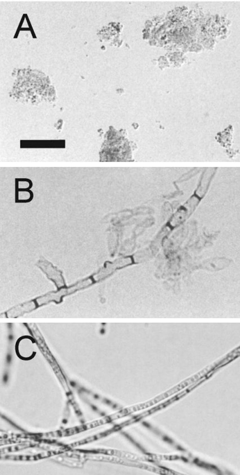
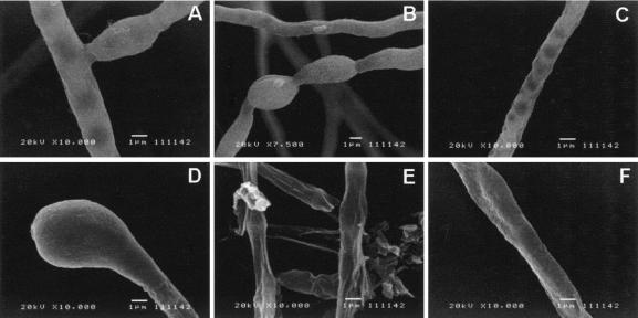
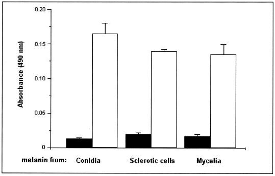
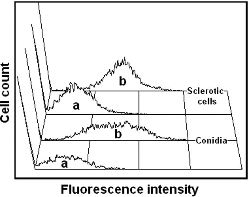
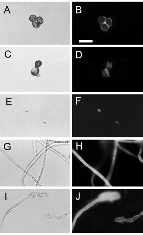
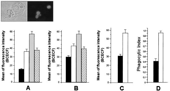
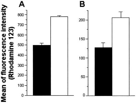
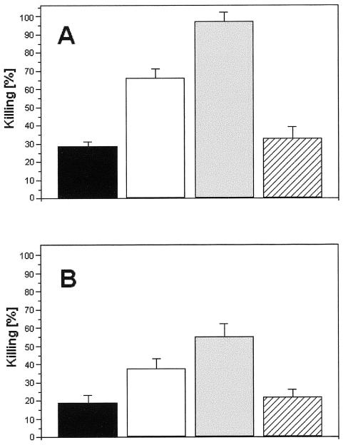
Fonsecaea pedrosoi is a fungal pathogen that produces melanin. The functions of melanin and its possible influence in the protective immunological response during infection by F. pedrosoi are not known. In this work, treatment of F. pedrosoi mycelia with proteases and glycosidases followed by a denaturing agent and hot concentrated acid left a black residue. Scanning electron microscopy demonstrated that this processed melanized residue resembled very closely the intact mycelium in shape and size. Melanin particles were also isolated from culture fluids of conidia or sclerotic forms of F. pedrosoi. Secreted melanins were reactive with sera from infected human patients, suggesting that F. pedrosoi synthesizes melanin in vivo. The antibodies against melanin were purified from patients' sera and analyzed by indirect immunofluorescence. They reacted with sclerotic cells from patients' lesions as well as with sclerotic bodies cultivated in vitro, conidia, mycelia, and digested residues. Treatment of F. pedrosoi with purified antibodies against melanin inhibited fungal growth in vitro. The interaction of F. pedrosoi with phagocytes in the presence of melanin resulted in higher levels of fungal internalization and destruction by host cells, which was accompanied by greater degrees of oxidative burst. Taken together, these results indicate that melanin from F. pedrosoi is an immunologically active fungal structure that activates humoral and cellular responses that could help the control of chromoblastomycosis by host defenses.


Figures

FIG. 1.
FIG. 1.
Light microscopy of F. pedrosoi melanins. Isolated melanin from conidial culture fluids forms amorphous aggregates (A). Treatment of mycelia with lytic enzymes and denaturing agents results in the production of black residues (B) that closely resemble intact mycelia (C). Bar, 10 μm.
FIG. 2.
FIG. 2.
SEM of F. pedrosoi viable mycelia (A to C) or digested preparations (D to F) (see Materials and Methods for details). As suggested by light microscopy, treatment of mycelia with lytic enzymes and denaturing agents results in the production of sediments that are very similar to intact cells in shape and size.
FIG. 3.
FIG. 3.
Antibodies against fungal melanin are produced during chromoblastomycosis. The reactivity of sera from healthy individuals (black bars) or patients with chromoblastomycosis (white bars) with melanin particles from culture fluids of conidia or sclerotic cells, as well as with cell-associated mycelial melanin, was tested by ELISA. The reactivity of patients’ sera was significantly higher than that of uninfected sera (P < 0.001). Results are expressed as means of three independent experiments ± standard deviations.
FIG. 4.
FIG. 4.
Purified antibodies against melanin bind conidia and sclerotic forms of F. pedrosoi. Fungal cells were incubated in the presence of human antibodies against melanin followed by FITC-labeled anti-human antibodies and were analyzed by flow cytometry. Fungal cells that had not been incubated with antibodies against melanin (a) showed basal levels of fluorescence, while fungal forms that were incubated with melanin antibodies (b) became fluorescent.
FIG. 5.
FIG. 5.
Cell wall distribution of melanin in F. pedrosoi. Sclerotic cells from patient lesions (A and B) or from in vitro cultivation (C and D), conidia (E and F), mycelia (G and H), or melanin ghosts (I and J) were incubated with antibodies against melanin followed by FITC-labeled anti-human antibodies and were analyzed by fluorescence microscopy. Left panels show fungal cells under differential interference contrast, while right panels show the same images under fluorescence. Bar, 10 μm.
FIG. 6.
FIG. 6.
Soluble melanin influences the ingestion of fungal cells by human or animal phagocytes. The indices of phagocytosis for F. pedrosoi conidia (A), sclerotic forms (B), or C. albicans organisms (C) by human neutrophils were expressed as a function of BCECF staining, as measured by flow cytometry. The interaction of F. pedrosoi conidia with mouse macrophages was evaluated microscopically after staining with Giemsa stain (D). Black bars, phagocytosis of fungal forms after interaction with phagocytes. The addition of soluble melanin (white bars) caused an increase in the phagocytosis levels in all systems. The influence of melanin antibodies alone (gray bars) or in association with melanin (hatched bars) on the phagocytosis of conidia (A) or sclerotic cells (B) was also evaluated. Values of P and statistical significance are described in Results. Data are expressed as means of three independent experiments ± standard deviations. Inset, representative sample of the interaction of BCECF-labeled conidia and human neutrophils.
FIG. 7.
FIG. 7.
Influence of soluble melanin in the oxidative burst of human neutrophils during incubation with F. pedrosoi conidia (A) or sclerotic bodies (B). The degrees of oxidative burst were measured by the emission of fluorescence after the oxidation of DHR to rhodamine. Bars represent the intensity of rhodamine fluorescence after interaction of F. pedrosoi with neutrophils in the absence (black bars) or presence (white bars) of soluble melanin. In the presence of soluble melanin, oxidative burst was significantly enhanced after interaction of neutrophils with conidia (P < 0.005) or sclerotic forms (P < 0.01). Results are expressed as means of three independent experiments ± standard deviations.
FIG. 8.
FIG. 8.
Killing of F. pedrosoi conidia (A) or sclerotic cells (B) after interaction with human neutrophils. Black bars, percentage of killing of fungal cells after interaction with neutrophils. Supplementation of the medium of interaction with melanin (white bars) or melanin antibodies (gray bars) significantly enhanced the antifungal efficacy of neutrophils, while the association between these components (hatched bars) resulted in levels of fungal death similar to those observed in the absence of antibodies or soluble melanin. Values of P and statistical significance are described in Results. Data are expressed as means of three independent experiments ± standard deviations.

References

    1. Alviano, C. S., S. R. Farbiarz, W. De Souza, J. Angluster, and L. R. Travassos. 1991. Characterization of Fonsecaea pedrosoi melanin. J. Gen. Microbiol. 137:837-844. - PubMed
    1. Alviano, C. S., L. R. Travassos, J. Angluster, and W. De Souza. 1992. Effect of environmental factors on Fonsecaea pedrosoi morphogenesis with emphasis on sclerotic cells induced by propranolol. Mycopathologia 119:17-23. - PubMed
    1. Blinova, M. I., N. M. Yudintseva, N. V. Kalmykova, E. V. Kuzminykh, N. A. Yurlova, O. A. Ovchinnikova, and I. L. Potokin. 2003. Effect of melanins from black yeast fungi on proliferation and differentiation of cultivated human keratinocytes and fibroblasts. Cell. Biol. Int. 27:135-146. - PubMed
    1. Butterfield, W., and S. C. Jong. 1987. Effect of carbon source on conidiogenesis in Fonsecaea dermatitidis, agent of chromomycosis. Mycopathologia 58:59-62. - PubMed
    1. D'Acquisto, F., R. Carnuccio, M. D'Ischia, and G. Misuraca. 1995. 5,6-Dihydroxyindole-2-carboxylic acid, a diffusible melanin precursor, is a potent stimulator of lipopolysaccharide-induced production of nitric oxide by J774 macrophages. Life Sci. 57:401-406. - PubMed

Publication types

LinkOut - more resources