. 2004 Jan;72(1):489-97.
doi: 10.1128/IAI.72.1.489-497.2004.

Listeriosis in the pregnant guinea pig: a model of vertical transmission

Affiliations

Listeriosis in the pregnant guinea pig: a model of vertical transmission

Anna I Bakardjiev et al. Infect Immun. 2004 Jan.

Abstract

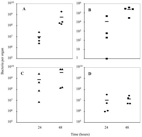
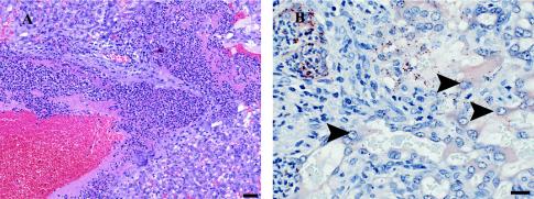
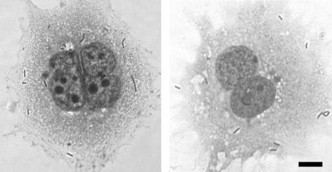
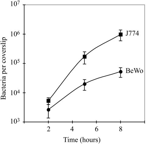
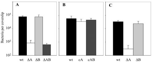
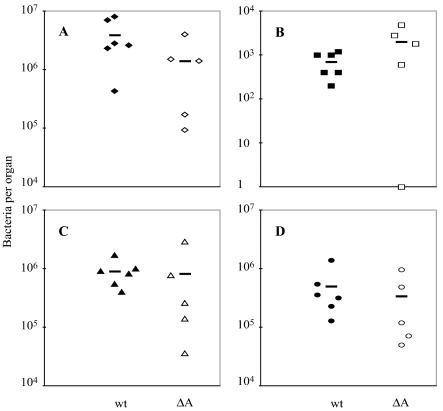
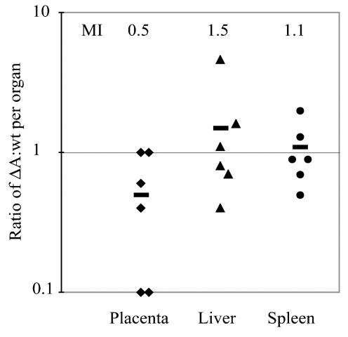
Feto-placental infections represent a major cause of pregnancy complications, and yet the underlying molecular and cellular mechanisms of vertical transmission are poorly understood. Listeria monocytogenes, a facultative intracellular pathogen, is one of a group of pathogens that are known to cause feto-placental infections in humans and other mammals. The purpose of this study was to evaluate possible mechanisms of vertical transmission of L. monocytogenes. Humans and guinea pigs have a hemochorial placenta, where a single layer of fetally derived trophoblasts separates maternal from fetal circulation. We characterized L. monocytogenes infection of the feto-placental unit in a pregnant guinea pig model and in primary human trophoblasts and trophoblast-derived cell lines. The clinical manifestations of listeriosis in the pregnant guinea pigs and the tropism of L. monocytogenes to the guinea pig placenta resembled those in humans. Trophoblast cell culture systems were permissive for listerial growth and cell-to-cell spread and revealed that L. monocytogenes deficient in internalin A, a virulence factor that mediates invasion of nonphagocytic cells, was 100-fold defective in invasion. However, crossing of the feto-placental barrier in the guinea pig model was independent of internalin A, suggesting a negligible role for internalin-mediated direct invasion of trophoblasts in vivo. Further understanding of vertical transmission of L. monocytogenes will help in designing more effective means of treatment and disease prevention.


Figures

FIG. 1.
FIG. 1.
Listeriosis in pregnant guinea pigs between days 42 and 52 of gestation. Bacteria per placenta (A), fetal liver (B), maternal liver (C), and maternal spleen (D) were enumerated at 24 or 48 h after intravenous inoculation of animals with 2 × 10⁷ wild-type L. monocytogenes organisms. The bars represent means.
FIG. 2.
FIG. 2.
Histological examination of guinea pig placental sections. Pregnant guinea pigs between days 42 and 52 of gestation were inoculated intravenously with 2 × 10⁷ wild-type L. monocytogenes bacteria. (A) Hematoxylin-eosin staining of placental section, showing the labyrinthine region at 24 h postinoculation. Moderate numbers of neutrophils and very small numbers of macrophages infiltrate the central region of the lobe. There is fibrin deposition associated within this infiltrate. Bar, 50 μm. (B) Immunohistochemistry for L. monocytogenes reveals large numbers of immunoreactive bacteria within the inflammatory infiltrate. Some bacteria appear to be inside trophoblasts (arrowheads). Bar, 20 μm.
FIG. 3.
FIG. 3.
Light micrographs of Diff-Quik stains of trophoblasts 2 h after infection with wild-type L. monocytogenes. (Left panel) Primary human trophoblasts. (Right panel) BeWo cells. In each case, gentamicin (50 μg/ml) was added 1 h after infection. Bar, 10 μm.
FIG. 4.
FIG. 4.
Growth of L. monocytogenes in BeWo cells and J774 murine macrophages. BeWo cells (3 × 10⁴) and J774 cells (6 × 10⁴) were plated on glass coverslips and grown overnight, leading to formation of monolayers. The difference in the number of plated BeWo and J774 cells reflects the difference in size between these two cell types. Cells were infected with a 1:20,000 dilution of an overnight culture of wild-type L. monocytogenes, resulting in MOIs of 1:8 for BeWo cells and 1:15 for J774 cells. At the specified times after infection, cells were lysed, and the number of bacteria per coverslip was determined in triplicate. In each case, gentamicin (50 μg/ml) was added 1 h after infection. Each growth curve represents the means and standard deviations from three separate experiments.
FIG. 5.
FIG. 5.
Invasion of trophoblasts by L. monocytogenes. Monolayers of cells were grown on glass coverslips. Trophoblasts were infected with L. monocytogenes 10403S (wild type [wt]) and the InlA deletion mutant (ΔA), the InlB deletion mutant (ΔB), the InlA InlB double deletion mutant (ΔAB), the InlA complementation mutant (cA), and the InlA InlB complementation mutant (cAB). BeWo cells were infected at an MOI of 1:1 (A and B), and primary human trophoblasts were infected at an estimated MOI of 20:1 (C). In each case, gentamicin (50 μg/ml) was added 1 h after infection. At 2 h after infection, monolayers were lysed and the number of bacteria per coverslip was determined in triplicate. All bacterial numbers are the averages and standard deviations from three separate experiments.
FIG. 6.
FIG. 6.
Infection of pregnant guinea pigs with wild-type L. monocytogenes (wt) or the InlA deletion mutant (ΔA). Groups of one to three pregnant guinea pigs between days 42 and 52 of gestation were inoculated intravenously with 106 bacteria. Bacteria per placenta (A), fetal liver (B), maternal liver (C), and maternal spleen (D) were enumerated at 48 h postinoculation. Bars represent means.
FIG. 7.
FIG. 7.
Competitive index analysis of InlA deletion mutant (ΔA) and wild-type (wt) L. monocytogenes. Groups of three pregnant guinea pigs were inoculated intravenously with a 1:1 mixture of the two strains at a total dose of 2 × 10⁷. Bacteria per organ were enumerated at 24 h postinoculation. Ratios between erythromycin-sensitive (ΔA) and erythromycin-resistant (wt) colonies were calculated for placenta, maternal liver, and spleen. Bars represent mean indices (MI).

References

    1. Auerbuch, V., L. L. Lenz, and D. A. Portnoy. 2001. Development of a competitive index assay to evaluate the virulence of Listeria monocytogenes actA mutants during primary and secondary infection of mice. Infect. Immun. 69:5953-5957. - PMC - PubMed
    1. Bishop, D. K., and D. J. Hinrichs. 1987. Adoptive transfer of immunity to Listeria monocytogenes. The influence of in vitro stimulation on lymphocyte subset requirements. J. Immunol. 139:2005-2009. - PubMed
    1. Braun, L., and P. Cossart. 2000. Interactions between Listeria monocytogenes and host mammalian cells. Microbes Infect. 2:803-811. - PubMed
    1. Cabanes, D., P. Dehoux, O. Dussurget, L. Frangeul, and P. Cossart. 2002. Surface proteins and the pathogenic potential of Listeria monocytogenes. Trends Microbiol. 10:238-245. - PubMed
    1. Camilli, A., L. G. Tilney, and D. A. Portnoy. 1993. Dual roles of plcA in Listeria monocytogenes pathogenesis. Mol. Microbiol. 8:143-157. - PMC - PubMed

Publication types

Substances