. 2004 Jan;16(1):201-14.
doi: 10.1105/tpc.014787. Epub 2003 Dec 19.

Maize mutants lacking chloroplast FtsY exhibit pleiotropic defects in the biogenesis of thylakoid membranes

Affiliations

Maize mutants lacking chloroplast FtsY exhibit pleiotropic defects in the biogenesis of thylakoid membranes

Yukari Asakura et al. Plant Cell. 2004 Jan.

Abstract

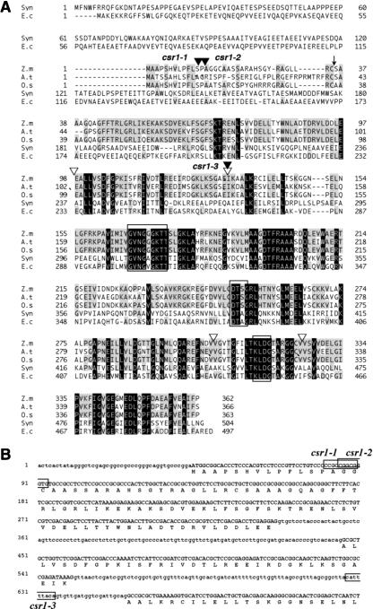
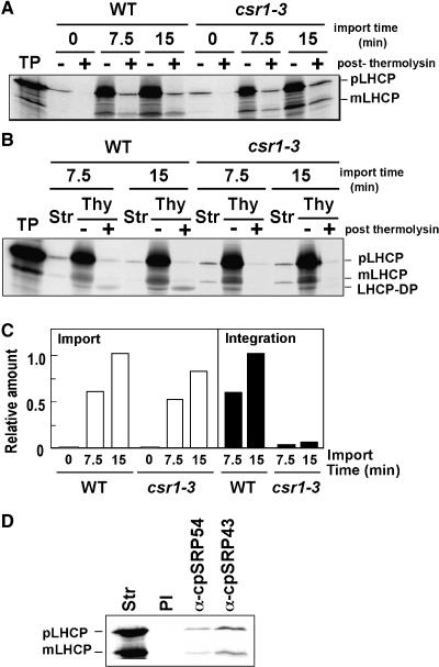
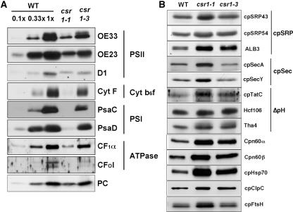
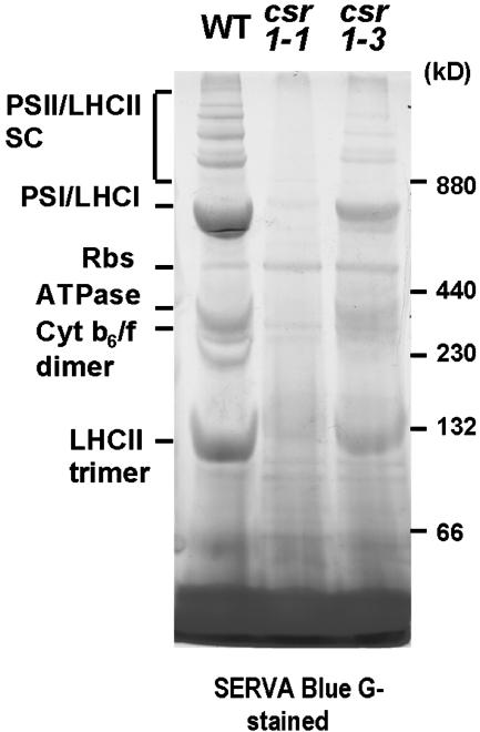
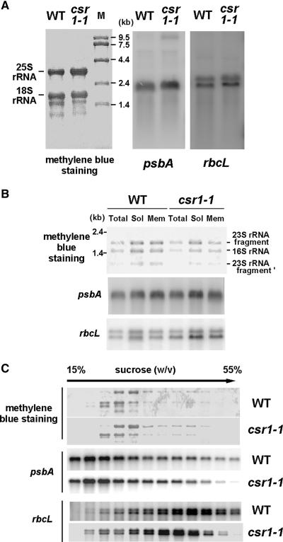
A chloroplast signal recognition particle (SRP) that is related to the SRP involved in secretion in bacteria and eukaryotic cells is used for the insertion of light-harvesting chlorophyll proteins (LHCPs) into the thylakoid membranes. A conserved component of the SRP mechanism is a membrane-bound SRP receptor, denoted FtsY in bacteria. Plant genomes encode FtsY homologs that are targeted to the chloroplast (cpFtsY). To investigate the in vivo roles of cpFtsY, we characterized maize cpFtsY and maize mutants having a Mu transposon insertion in the corresponding gene (chloroplast SRP receptor1, or csr1). Maize cpFtsY accumulates to much higher levels in leaf tissue than in roots and stems. Interestingly, it is present at similar levels in etiolated and green leaf tissue and was found to bind the prolamellar bodies of etioplasts. A null cpFtsY mutant, csr1-1, showed a substantial loss of leaf chlorophyll, whereas a "leaky" allele, csr1-3, conditioned a more moderate chlorophyll deficiency. Both alleles caused the loss of various LHCPs and the thylakoid-bound photosynthetic enzyme complexes and were seedling lethal. By contrast, levels of the membrane-bound components of the thylakoid protein transport machineries were not altered. The thylakoid membranes in csr1-1 chloroplasts were unstacked and reduced in abundance, but the prolamellar bodies in mutant etioplasts appeared normal. These results demonstrate the essentiality of cpFtsY for the biogenesis not only of the LHCPs but also for the assembly of the other membrane-bound components of the photosynthetic apparatus.


Figures

Figure 1.
Figure 1.
Deduced Amino Acid Sequence of Maize cpFtsY and Corresponding Positions of Mu Insertions in csr1 Mutants. (A) The maize cpFtsY sequence (Z.m) was aligned with those of chloroplast homologs from Arabidopsis thaliana (A.t) and Oryza sativa (O.s) and also with bacterial counterparts in Synechocystis PCC6803 (Syn) and Escherichia coli (E.c). Residues that are identical among all of the aligned proteins are shaded in black. Residues shared by at least three of the proteins are shaded in gray. Three consensus motifs for a GTP binding domain are boxed. A predicted cleavage site by stromal processing protease in maize cpFtsY is indicated by the arrow. Corresponding positions of Mu transposon insertions in the csr1-1, csr1-2, and csr1-3 alleles are indicated by closed triangles, and those of known introns in the genomic sequence are indicated by open triangles. (B) Partial nucleotide sequence of csr1 and sites of Mu insertions. The sequence shown begins 43 nucleotides upstream of the start codon and ends within the third exon. The deduced amino acid sequence is indicated below. Locations of Mu insertions are shown by boxes to indicate the sequences duplicated upon insertion. csr1-1 contains a MuDR insertion, whereas csr1-2 and csr1-3 carry Mu1 insertions.
Figure 2.
Figure 2.
Immunoblot Analyses of Maize cpFtsY: Tissue and Light Dependence, and Suborganellar Localization. (A) Abundance of cpFtsY in different maize tissues. Twenty micrograms each of total protein extracted from roots, stems, and leaves of light-grown maize seedlings were analyzed by immunoblotting with anti-cpFtsY antibodies (top gel) or by Coomassie blue staining (bottom gel). (B) Abundance of cpFtsY in dark-grown and light-grown leaves. Maize seedlings were grown either for 9 days in the dark (D) or for 10 days in the light (L). Dark-grown seedlings were grown further in the light for the indicated times (2, 6, 12, 24, and 48 h). Leaf extracts were prepared and examined by immunoblot analysis with the indicated antibodies. (C) Suborganellar localization of cpFtsY. In the gels at left, chloroplasts (Chl) were fractionated into the envelope membranes (Env), the stroma (Str), and the thylakoids (Thy). Isolated thylakoids were treated with 30 μg/mL trypsin on ice for 5 min. Equivalent amounts of each fraction were examined by immunoblot analysis. In the gels at right, etioplasts were fractionated as described in Methods and analyzed as described for the gels at left. The asterisks denote a characteristic proteolytic fragment derived from ALB3. Pro, prolamellar body.
Figure 3.
Figure 3.
Pigment Deficiency of csr1 Mutant Seedlings. (A) Wild-type (WT), csr1-1, and csr1-3 seedlings were photographed after 10 days of growth under the conditions described in Methods. (B) Chlorophyll content of csr1 mutant leaves. Chlorophyll was extracted from the leaves of 10-day-old seedlings and quantified as described in Methods. Average values obtained from two independent experiments (n = 4 or 5 each) are plotted per 100 mg fresh weight (FW). (C) Immunoblot analysis of leaf cpFtsY in the mutant seedlings. Leaf proteins (20 μg) of wild-type, csr1-1, and csr1-3 seedlings were electrophoresed by 12.5% SDS-PAGE and analyzed using anti-maize cpFtsY IgGs.
Figure 4.
Figure 4.
Ultrastructure of Chloroplasts and Etioplasts in csr1 Mutants. (A) Reduced thylakoid membrane content in csr1 mutant chloroplasts. WT, wild type. (B) Reduced prothylakoid membranes in csr1-1 mutant etioplasts. Bars = 500 nm.
Figure 5.
Figure 5.
LHCP Immunoblot Analyses of csr1 Mutants. Immunoblot analyses of individual members of the LHCP family in wild-type (WT), csr1-1, and csr1-3 leaf extracts. Extracts were the same as those shown in Figure 3C. Two micrograms of protein was analyzed for Lhca1, Lhca2, Lhca3, Lhca4, Lhcb1, Lhcb2, and Lhcb6. Six micrograms of protein was analyzed for Lhcb5, and 20 μg of protein was analyzed for Lhcb3. Dilutions of 0.33× and 0.1× of the wild-type extract also were analyzed to aid in quantification.
Figure 6.
Figure 6.
In Organello Import and Membrane Insertion of LHCP in csr1 Mutant Chloroplasts. Intact chloroplasts isolated from wild-type or csr1-3 leaves were incubated with in vitro–synthesized pLHCP in a chloroplast import reaction as described in Methods. Fluorograms after SDS-PAGE are shown. (A) pLHCP import in wild-type (WT) and csr1-3 chloroplasts. Import reactions were incubated for 0, 7.5, or 15 min as indicated. Postimport thermolysin treatments (post thermolysin) were performed at 100 μg/mL for 20 min on ice. Mature LHCP (mLHCP) was not detected in the 0-min samples. TP, in vitro translation products. (B) Insertion of LHCP into the thylakoid membranes assessed by postimport thermolysin treatment of thylakoid membranes. After a 7.5- or 15-min import reaction, reaction products were placed on ice and chloroplasts were recovered and fractionated into stroma (Str) and thylakoids (Thy). Proper LHCP insertion into thylakoids was analyzed by treating the thylakoids with thermolysin at 100 μg/mL for 40 min on ice (post thermolysin). LHCP-DP is a thermolysin-resistant LHCP fragment that reflects the correct insertion of LHCP into thylakoid membranes (Cline, 1986). (C) Quantitative analysis of the data shown in (A) and (B). The amounts of thermolysin-resistant mLHCP after import (A) and of LHCP-DP in the thermolysin-treated thylakoids (B) were quantified densitometrically and are indicated as relative amounts of LHCP import into the chloroplasts (left graph) and proper integration into the thylakoid membranes (right graph), respectively. In both cases, the amount of thermolysin-resistant mLHCP or LHCP-DP after a 15-min import into the wild type was set to 1. (D) Association of newly imported LHCP with cpSRP components in csr1-3 mutant stroma. Import reactions with csr1-3 chloroplasts were incubated for 30 min and fractionated to obtain the stromal fraction (Str). Fifty-microliter aliquots were immunoprecipitated with 20 μL of anti-cpSRP54, anti-cpSRP43, or preimmune (PI) IgGs as described in Methods. An equivalent fraction of each immunoprecipitate was analyzed.
Figure 7.
Figure 7.
Immunoblot Analyses of Photosynthetic Thylakoid Membrane Proteins, Stromal Chaperones, and Components of the Thylakoid Protein Translocation Machineries in csr1 Mutants. Total leaf proteins prepared from wild-type, csr1-1, and csr1-3 seedlings were examined by immunoblot analysis with the indicated antibodies. Extracts were the same as those used in Figure 3C. (A) Immunoblot analyses of photosynthetic thylakoid membrane proteins. The complex to which each protein belongs is indicated. Two micrograms of leaf protein was analyzed for OE33, CytF, PsaC, CF1α, and CFoI. Six micrograms of leaf protein was analyzed for D1, and 20 μg of leaf protein was analyzed for OE23 and PsaD. Dilutions of 0.33× and 0.1× of wild-type (WT) extracts also were analyzed for comparison. (B) Immunoblot analysis of components of the cpSRP, cpSec, and ΔpH thylakoid import machineries and of stromal chaperone proteins. Two micrograms of total leaf protein was analyzed for cpHsp70, Cpn60α, and Cpn60β. Five micrograms of total leaf protein was analyzed for ALB3. Ten micrograms of total leaf protein was analyzed for cpFtsH, Hcf106, and Tha4. Twenty micrograms of total leaf protein was analyzed for cpSRP54, cpSRP43, cpSecY, and cpTatC.
Figure 8.
Figure 8.
Photosynthetic Complexes Were Decreased Dramatically in the csr1 Mutants. Chloroplast proteins (48 μg) from wild type (WT), csr1-1, and csr1-3 seedling leaves were solubilized by treatment with 1% (w/v) n-dodecyl-β-d-maltoside and separated by 5 to 14% blue native gel electrophoresis. Without additional staining, protein complexes were detected by bound Coomassie blue dye (SERVA Blue G-stained) and chlorophylls. Bands corresponding to various photosynthetic complexes in wild-type seedling leaves are indicated and were identified by immunoblot analysis, as shown in the supplemental data online (see also http://www.hos.ufl.edu/clineweb/BNgel.htm). PSII/LHCII SC, PSII/LHCII supercomplex; PSI/LHCI, PSI and LHCI; Rbs, ribulose-1,5-bisphosphate carboxylase/oxygenase. Ferritin (880 and 440 kD), catalase (230 kD), and BSA (132 and 66 kD) were used as molecular mass marker proteins.
Figure 9.
Figure 9.
Abundance, Membrane Association, and Polysome Association of the psbA mRNA Are Not Affected Significantly in the csr1-1 Mutant. (A) Total leaf RNA (5 μg) was analyzed by RNA gel blot hybridization using a radiolabeled psbA fragment (center gel) or rbcL fragment (right gel). The same samples were analyzed by methylene blue staining to visualize rRNAs (left gel). The positions of cytosolic 25S rRNA and 18S rRNA are indicated. WT, wild type. (B) RNAs were purified from the membrane (Mem) and soluble (Sol) fractions of wild-type or csr1-1 chloroplasts as described in Methods. Equivalent proportions of the membrane-bound and soluble RNAs, and one-third of the RNA prepared from the corresponding starting chloroplast sample, were analyzed by methylene blue staining (top gel) or by hybridization using the psbA (middle gel) or rbcL (bottom gel) probe. The positions of chloroplast 16S rRNA and 23S rRNA fragments are indicated. (C) Analysis of leaf polysomes in the csr1-1 mutant. Total leaf extracts were prepared under conditions that maintained the integrity of polysomes and fractionated in 15 to 55% (w/v) sucrose gradients. RNAs were prepared from each fraction and examined by RNA gel blot analysis as described for (A).

References

    1. Allen, J.F., and Forsberg, J. (2001). Molecular recognition in thylakoid structure and function. Trends Plant Sci. 6, 317–326. - PubMed
    1. Amin, P., Sy, D.A., Pilgrim, M.L., Parry, D.H., Nussaume, L., and Hoffman, N.E. (1999). Arabidopsis mutants lacking the 43- and 54-kilodalton subunits of the chloroplast signal recognition particle have distinct phenotypes. Plant Physiol. 121, 61–70. - PMC - PubMed
    1. Andersson, J., Walters, R.G., Horton, P., and Jansson, S. (2001). Antisense inhibition of the photosynthetic antenna proteins CP29 and CP26: Implications for the mechanism of protective energy dissipation. Plant Cell 13, 1193–1204. - PMC - PubMed
    1. Arnon, D.I. (1949). Copper enzymes in isolated chloroplasts. Plant Physiol. 24, 1–15. - PMC - PubMed
    1. Aronsson, H., Sundqvist, C., and Dahlin, C. (2003). POR: Import and membrane association of a key element in chloroplast development. Physiol. Plant. 118, 1–9. - PubMed

Publication types

MeSH terms

Substances

Associated data