

Evaluating requirements for spatial resolution of fMRI for neurosurgical planning
- PMID: 14689508
- PMCID: PMC6872071
- DOI: 10.1002/hbm.10148
Evaluating requirements for spatial resolution of fMRI for neurosurgical planning
Abstract
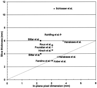
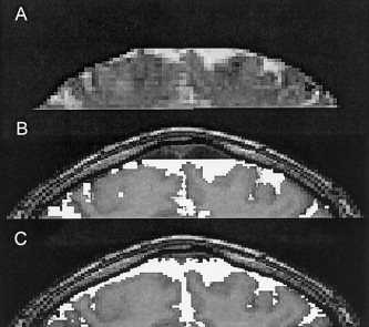
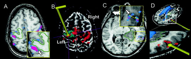
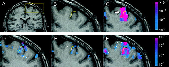
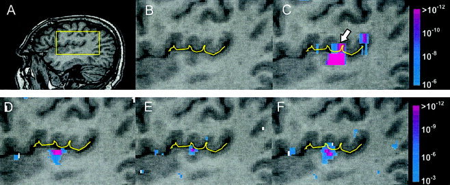
The unambiguous localization of eloquent functional areas is necessary to decrease the neurological morbidity of neurosurgical procedures. We explored the minimum spatial resolution requirements for functional magnetic resonance imaging (fMRI) data acquisition when brain mapping is used in neurosurgical planning and navigation. Using a 1.5 Tesla clinical MRI scanner, eight patients with brain tumors underwent fMRI scans using spatial resolution of approximately 4 x 4 x 4 mm(3) to map the eloquent motor and language areas during the performance of cognitive/sensorimotor tasks. The fMRI results were then used intra-operatively in an open MRI system to delineate eloquent areas. Retrospectively, activation patterns were visually inspected by a neurosurgeon to determine qualitatively whether ambiguity with respect to the activation boundaries, due to low spatial resolution, could be of potential significance for surgical guidance. A significant degree of ambiguity in both the extent and shape of activation was judged to be present in data from six of the eight patients. Analysis of fMRI data at multiple resolutions from a normal volunteer showed that at 3 mm isotropic resolution, eloquent areas were better localized within the gray matter although there was still some potential for ambiguity caused by activations appearing to cross a sulcus. The data acquired with 2-mm isotropic voxels significantly enhanced the spatial localization of activation to within the gray matter. Thus, isotropic spatial resolution on the order of 2 x 2 x 2 mm(3), which is much higher than the resolutions used in typical fMRI examinations, may be needed for the unambiguous identification of cortical activation with respect to tumors and important anatomical landmarks.
Copyright 2003 Wiley-Liss, Inc.
Figures
References
-
- Berger MS, Deliganis AV, Dobbins J, Keles GE (1994): The effect of extent of resection on recurrence inxpatients with low grade cerebral hemisphere gliomas. Cancer 74: 1784–1791. - PubMed
-
- Bittar RG, Olivier A, Sadikot AF, Andermann F, Pike GB, Reutens DC (1999): Presurgical motor and somatosensory cortex mapping with functional magnetic resonance imaging and positron emission tomography. J Neurosurg 91: 915–921. - PubMed
-
- Chen NK, Dickey CC, Yoo SS, Guttmann CR, Panych LP (2003): Selection of voxel size and slice orientation for fMRI in the presence of susceptibility field gradients: application to imaging of the amygdala. Neuroimage 19: 817–825. - PubMed
-
- Cifelli A, Matthews PM (2002): Cerebral plasticity in multiple sclerosis: insights m sixfMRI. Mult Scler 8: 193–199. - PubMed
Publication types
MeSH terms
Grants and funding
LinkOut - more resources
Full Text Sources
Medical
