Review
. 2004 Jan;204(1):13-24.
doi: 10.1111/j.1469-7580.2004.00260.x.

Identifying genes for neuron survival and axon outgrowth in Hirudo medicinalis

Affiliations
Review

Identifying genes for neuron survival and axon outgrowth in Hirudo medicinalis

S E Blackshaw et al. J Anat. 2004 Jan.

Abstract

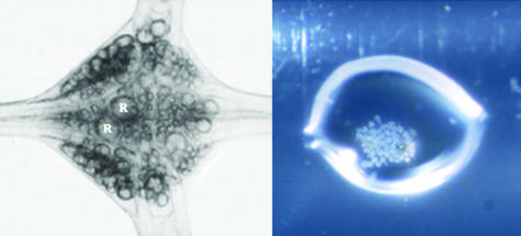
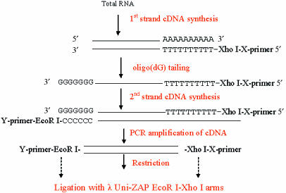
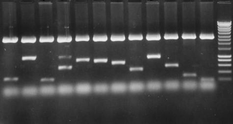
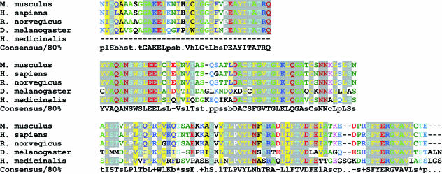
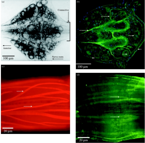
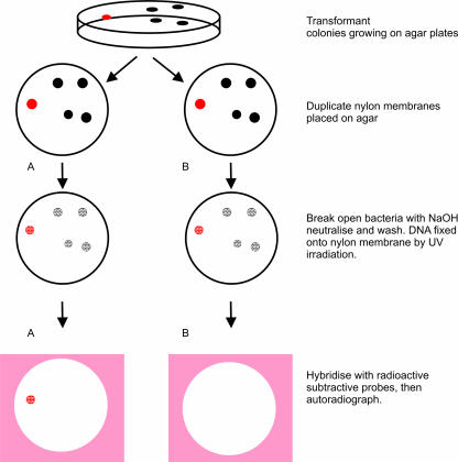
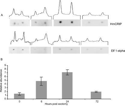
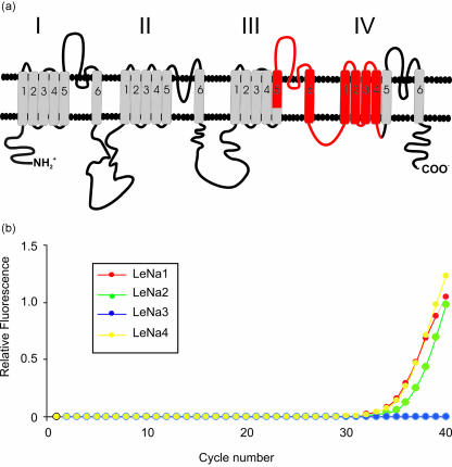
We have studied the molecular basis of nervous system repair in invertebrate (Hirudo medicinalis) nerve cells. Unlike in mammals, neurons in invertebrates survive injury and regrow processes to restore the connections that they held before the damage occurred. To identify genes whose expression is regulated after injury, we have used subtractive probes, constructed from regenerating and non-regenerating ganglia from the leech Hirudo medicinalis, to screen cDNA libraries made from whole leech CNS or from identified microdissected neurons. We have identified genes of known or predicted function as well as novel genes. Known genes up-regulated within hours of injury and that are widely expressed in invertebrate and mammalian cells include thioredoxin and tubulin. Other known genes, e.g. Cysteine Rich Intestinal Protein (CRIP), have previously been identified in mammalian cells though not in regenerating adult neurons. Two regulated genes identified, myohemerythrin and the novel protein ReN3 are exclusively expressed in invertebrates. Thus our approach has enabled us to identify genes, present in a neuron of known function, that are up- and down-regulated within hours of axotomy, and that may underpin the intrinsic ability of invertebrate neurons to survive damage and initiate regrowth programmes.


Figures

Fig. 1
Fig. 1
cDNA libraries were constructed using whole ganglia from the leech ventral nerve cord or microdissected neuron cell bodies. (A) An isolated ganglion contains around 400 nerve cells including primary sensory and motor neurons, modulatory neurons and interneurons, together with macroglia and microglia, and the cells comprising the external connective tissue sheath, and the internal capsule, a structure which separates the cell bodies from the central neuropile area of the ganglion. R, Retzius cells. (B) Isolated Retzius cell bodies microdissected from successive ganglia in the ventral nerve cord, collected into a drop of fluid on the wall of an Eppendorf tube, and used to construct the Retzius neuron library.
Fig. 2
Fig. 2
Scheme for library construction: flow chart of the construction of amplification-based cDNA libraries from single ganglia or microdissected neurons (modified from Korneev et al. 1996).
Fig. 3
Fig. 3
Retzius neuron library constructed using around 30 microdissected cell bodies. (A) Twelve clones picked at random from the library are illustrated on the agarose gel. Of the 12 illustrated clones, the average insert size was about 600 bp. Most of the clones had open reading frames and some clones had polyadenylation signals.
Fig. 4
Fig. 4
CLUSTAL alignment of the amino acid sequence of part of cytoplasmic dynein heavy chain in leech with homologous sequences from genomes of human, fly, mouse and rat.
Fig. 5
Fig. 5
Expression of the novel peptide, designatd ReN3, in nerve cells, connective tissue and muscle of adult leech CNS. ReN3 is one of four novel clones isolated to date from the 24-h regenerating ganglion library. It has no known homologue in vertebrate genomes. (A) Isolated segmental ganglion viewed under transmitted light; ventral surface. The boxed area of the connectives is shown in C and D. (B) Horizontal section through the ganglion; orientation as in A. Immunohistochemistry using an antibody to a custom peptide shows that the encoded ReN3 peptide is widely expressed in neuron cell bodies (long arrows), in the connective tissue capsule that surrounds the ganglion (short arrows) and in the internal capsule of the ganglion (long arrows). The section is counterstained with DAPI to show nuclei. (C,D) ReN3 is also expresed in the muscle cells that extend throughout the ventral nerve cord within the connective tissue capsule. (C) A confocal section through part of the connectives that link adjacent ganglia of the ventral nerve cord (see boxed area in A), in a preparation stained with rhodamine–phalloidin to illustrate the muscle layout. (D) A confocal section through an equivalent region stained with the ReN3 antibody.
Fig. 6
Fig. 6
Protocol used to screen differentially the Retzius neuron library to identify clones regulated 24 h after injury and which are also expressed in this serotonergic neuron. Clones from the library were transferred in bulk to nylon membranes. Duplicate plaque lifts from 90-mm plates are shown diagrammatically. Two copies of the clones were made (A,B) to allow hybridization with the control and 24-h regenerating probes. This diagram illustrates screening of five clones: clones from the Retzius library that have hybridized with the control probe in A and with the 24-h regenerating probe in B. Comparing these autoradiographs identifies clones that are differentially expressed. For example, the red spot in A indicates a clone that is down-regulated after axotomy, denoted by the positive hybridization signal on the control film, not present on the 24-h film. The autoradiograph was then used to identify the relevant clone on the agar colony and these were excised from the agar, the DNA purified and sent for sequencing.
Fig. 7
Fig. 7
Semiquantitative expression of Cysteine Rich Intestinal Protein (CRIP): filters were dotted with spots of two genes, CRIP and Elongation Factor 1-α. The filters were probed with radiolabelled cDNA produced from five ganglia at chosen time points. Specific activity is measured using an NCBI image programme that determines how dark each spot was. This gives an assessment of the degree of hybridization, and hence a measure of expression level.
Fig. 8
Fig. 8
Identification of genes encoding voltage-gated sodium channel isoforms in single leech neurons. (A) Cartoon of the α-subunit of a voltage-gated Na channel representing the phospholipid bilayer; and the four domains of the channel labelled I–IV. Each of these domains has six probable transmembrane helices represented by the cylinders. The four domains come together in three dimensions to form the pore of the channel. To isolate Na-channel genes in Hirudo, we designed degnerate primers by comparing the amino acid sequence of the α-subunit of a rat brain (type IIa) Na-channel with published sequences of the putative channel in a range of different invertebrates (sea slug, fruit fly, squid, flatworm and jellyfish), and identified regions in domains III and IV of the α-subunit where the amino acid sequence is highly conserved. These primers span a segment of the α-ubunit, shown in red, 900 nucleotides long, between segment S5 of domain III and segment S4 of domain IV. This approach has isolated four homologous yet distinct isoforms of the leech Na-channel, designated LeNa 1–4. (B) Analysis of isoform expression in single Retzius neurons using real-time (quantitative) PCR shows that Retzius cells express multiple isoforms of the channel, LeNa isoforms 1, 2 and 4, but not isoform 3.

References

    1. Bannatyne BA, Blackshaw SE, McGregor MS. New growth elicited in adult leech mechanosensory neurones by peripheral axon damage. J. Exp. Biol. 1989;143:419–434. - PubMed
    1. Baylor DA, Nicholls JG. Patterns of regeneration between individual nerve cells in the central nervous system of the leech. Nature. 1971;232:268–270. - PubMed
    1. Birkenmeier EH, Gordon JI. Developmental regulation of a gene that encodes a cysteine-rich intestinal protein and maps near the murine immunoglobulin heavy chain locus. Proc. Natl Acad. Sci. USA. 1986;83:2516–2520. - PMC - PubMed
    1. Blackshaw SE, Gross RH, Porter DM, Henderson LP, Maue RA. Cloning of a family of putative sodium channel α subunit cDNA's from the leech. J. Physiol. 1999;518P:126.
    1. Blackshaw SE, Henderson LP, Malek J, Porter DM, Gross RH, Angstadt JD, et al. Single-cell analysis of a novel family of voltage-dependent sodium channel genes in the leech demonstrates cell-specific patterns of expression. J. Neurobiol. 2003;55:355–371. - PubMed

Publication types

LinkOut - more resources