Review
. 2004 Jan;204(1):25-31.
doi: 10.1111/j.1469-7580.2004.00262.x.

Factors secreted by Schwann cells stimulate the regeneration of neonatal retinal ganglion cells

Affiliations
Review

Factors secreted by Schwann cells stimulate the regeneration of neonatal retinal ganglion cells

Jeremy S H Taylor et al. J Anat. 2004 Jan.

Abstract

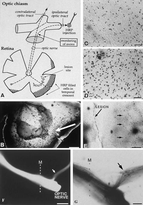
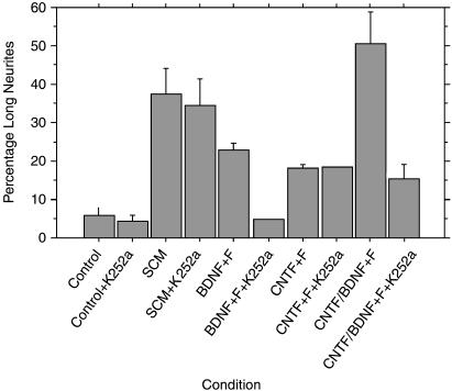
The adult mammalian central nervous system (CNS) does not repair after injury. However, we and others have shown in earlier work that the neonatal CNS is capable of repair and importantly of allowing regenerating axons to re-navigate through the same pathways as they did during development. This phase of neonatal repair is restricted by the fragility of neurons after injury and a lack of trophic factors that enable their survival. Our aim is to define better the factors that sustain neurons after injury and allow regeneration to occur. We describe some of our work using Schwann cells to promote the regeneration of neurons from young postnatal rodents. We have established rapid methods for purifying Schwann cells without the use of either anti-mitotic agents to suppress contaminating fibroblasts or mitotic stimulation to generate large numbers of Schwann cells. The rapidly purified Schwann cells have been used to generate conditioned medium that we have shown stimulates axon regeneration in cultured retinal ganglion cell neurons. We also show that the positive effects of Schwann cells are still present after pharmacological blockade of the neurotrophin receptors, suggesting that novel factors mediate these effects.


Figures

Fig. 1
Fig. 1
Lesions of the marsupial (Monodelphis domestica) retina during the neonatal period result in regeneration with correct re-navigation of the visual pathways. (A) Schematic diagram showing the lesion of the temporo-ventral retina and position of HRP labelling in the ipsilateral optic tract, which was used to define the successful regeneration of RGCs with uncrossed projections. (B) The retina 10 days after lesion showing retrogradely labelled cells in the periphery behind the lesion (arrow). (C) Higher magnification view of regenerated retrogradely labelled retinal ganglion cells, which are fewer in number than those retrogradely labelled in un-lesioned control retinae (D). (E) The lesion and the regenerated RGCs. (F, G) Axons labelled with Dil (1, 1′ -dioctadecyl -3, 3, 3′, 3′ -tetramethylindocarbocyanide perchlorate) and photo-converted Dil, respectively, show the anterograde tracing of regenerating axons from behind a lesion in the temporo-ventral retina showing successful re-navigation of the optic chiasm. Scale bars in B and G, 200 µm; C and D, 25 µm; E, 50 µm; F, 500 µm.
Fig. 2
Fig. 2
Purified rat P4 Schwann cells 3 days after immunopanning stained with S100. Retinal ganglion cells isolated from P4 rat retina, stained with carboxyfluorescein diacetate, growing in the presence of Schwann cell conditioned medium. The extensive neurite outgrowth of the neurons is regenerative. Scale bars, 50 µm.
Fig. 3
Fig. 3
Graphs showing the effects of different Schwann cell conditioned media (SCM) on RGC regeneration. Positive control medium containing BDNF or a combination of CNTF/BDNF and forskolin have slightly better neuritogenic effects than SCM. The effects of BDNF, but not those of CNTF (which does not act through a Trk receptor), are lost when the kinase inhibitor K252a is added. By contrast, SCM shows minimal loss of function when K252a is added. This suggests that the SCM positive regeneration-promoting factors are not acting through neurotrophin receptors.
Fig. 4
Fig. 4
(A,B) A method used to radiolabel proteins made by Schwann cells, which are then fed to RGCs. The Schwann-cell-derived factors are taken up by RGCs and then isolated by fluorography. (B) Radiolabelled proteins in Schwann cell conditioned medium (lane 1) and after passing through a spin column to remove unbound radiolabel (lane 2). Lanes 3 and 4 show the residual radiolabelled SCM after incubation with retinal ganglion cells (different preparations). Lanes 5 and 6 show two different exposures of RGC lysates after 24 h incubation, showing a number of radiolabelled bands, including a clear band at 40 kDa (indicated by arrow), which has been taken up by RGCs from the Schwann cell conditioned medium.

References

    1. Bampton ETW, Taylor JSH. RT-PCR analysis of neurotrophic factor expression in Schwann cells prepared by different methods. Soc. Neurosci. 2001;27:219.16.
    1. Barres BA, Silverstein BE, Corey DP, Chun LL. Immunological, morphological, and electrophysiological variation among retinal ganglion cells purified by panning. Neuron. 1988;1:791–803. - PubMed
    1. Bermingham JR, Jr, Shumas S, Whisenhunt T, Rosenfeld MG, Scherer SS. Modification of representational difference analysis applied to the isolation of forskolin-regulated genes from Schwann cells. J. Neurosci. Res. 2001;63:516–524. - PubMed
    1. Berry M, Carlile J, Hunter A. Peripheral nerve explants grafted into the vitreous body of the eye promote the regeneration of retinal ganglion cell axons severed in the optic nerve. J. Neurocytol. 1996;25:147–170. - PubMed
    1. Brockes JP, Fields KL, Raff MC. Studies on cultured rat Schwann cells. I. Establishment of purified populations from cultures of peripheral nerve. Brain Res. 1979;165:105–118. - PubMed

MeSH terms

LinkOut - more resources