Review
. 2004 Jan;204(1):57-67.
doi: 10.1111/j.1469-7580.2004.00257.x.

Olfactory ensheathing cells (OECs) and the treatment of CNS injury: advantages and possible caveats

Affiliations
Review

Olfactory ensheathing cells (OECs) and the treatment of CNS injury: advantages and possible caveats

Susan C Barnett et al. J Anat. 2004 Jan.

Abstract

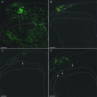
One of the main research strategies to improve treatment for spinal cord injury involves the use of cell transplantation. This review looks at the advantages and possible caveats of using glial cells from the olfactory system in transplant-mediated repair. These glial cells, termed olfactory ensheathing cells (OECs), ensheath the axons of the olfactory receptor neurons. The primary olfactory system is an unusual tissue in that it can support neurogenesis throughout life. In addition, newly generated olfactory receptor neurons are able to grow into the CNS environment of the olfactory bulb tissue and reform synapses. It is thought that this unique regenerative property depends in part on the presence of OECs. OECs share some of the properties of both astrocytes and Schwann cells but appear to have advantages over these and other glial cells for CNS repair. In particular, OECs are less likely to induce hypertrophy of CNS astrocytes. As well as remyelinating demyelinated axons, OEC grafts appear to promote the restoration of functions lost following a spinal cord lesion. However, much of the evidence for this is based on behavioural tests, and the mechanisms that underlie their potential benefits in transplant-mediated repair remain to be clarified.


Figures

Fig. 1
Fig. 1
Longitudinal section through the rat head. The olfactory system is a mixture of PNS and CNS tissue. After injury or during normal cell turnover, new olfactory receptor nerves (ORNs) are generated from basal stem cells in the olfactory epithelium located in the PNS. Throughout life these neurons are able to extend axons from the PNS, enter the CNS environment of the olfactory bulb and reform synapses with the mitral cells. (Modified from H. G. Liebich.)
Fig. 2
Fig. 2
Comparison of the properties of OECs and Schwann cells. Rat OECs and Schwann cells share: (1) antigenic and morphological characteristics – namely a spindle shape and express p75NTR, GFAP and O4 immunoreactivity (Barnett et al. 1993b; Mirsky & Jessen, 1999); (2) growth factor response to GGF, forskolin and FGF2 (Yan et al. 2001; Alexander et al. 2002); (3) connexin expression and gap junctional communication (Barnett et al. 2001); (4) transcriptional regulation during myelination in vivo (Smith et al. 2001). Rat OECs and Schwann cells may have differences in: (1) erbB receptor expression – Schwann cells express erbB2 and erbB3, and OECs express erbB2 and erbB4 (Thompson et al. 2000); (2) interaction with astrocytes – Schwann cells but not OECs induce hypertrophy in astrocytes as assessed by an increase in astrocyte cytoplasmic area and expression of chondroitin sulphate proteoglycans at the graft site (Lakatos et al. 2000; Plant et al. 2001).
Fig. 3
Fig. 3
Differences in erbB receptor profiles in OECs and Schwann cells. (A) RT-PCR illustrating olfactory bulb tissue (OB) and primary cultures of Schwann cells express the mRNA for erbB3. Conversely, OB and primary cultures of OECs but not Schwann cells express the mRNA for erbB4. M, molecular weight markers. (B) Immunofluorescence of OECs labelled with antibodies to erbB2, erbB3 and erbB4. These data show OECs express erbB2 and erbB4. Magnification ×200.
Fig. 4
Fig. 4
Schematic representation of the interaction of OECs (pink) and Schwann cells (red) with astrocytes in co-culture. The main difference in characteristics between OECs and Schwann cells is seen in their interactions with astrocytes (green). (A) Schwann cells do not mingle well and astrocytes express characteristics of hypertrophy. (B) Conversely, OECs mingle well among astrocytes and do not appear to evoke any changes in astrocytes.
Fig. 5
Fig. 5
Schematic diagrams illustrating the normal dorsal root entry zone and different regeneration scenarios. (A) Sensory axons enter the spinal cord at the dorsal root entry zone. In the spinal cord, afferent fibres give rise to branches that travel in the dorsal columns and axon collaterals that synapse with spinal cord neurons in the grey matter. (B) When dorsal roots are lesioned, the central axons of afferent fibres regenerate along the dorsal root until they reach the dorsal root entry zone where regeneration is abortive. Grafts of OECs are reported to allow regenerating dorsal root fibres to enter the spinal cord (C), and re-establish functional connections with spinal cord neurons (D).
Fig. 6
Fig. 6
Schematic diagram to illustrate plan of electrophysiological experiments to investigate the effects of OECs on regeneration at the dorsal root entry zone. (A) A lumbar dorsal root is sectioned and re-anastomosed, and in the same operation OECs are injected into the dorsal root and dorsal root entry zone. A loop of suture thread identifies the lesioned root. (B) Three to 6 months later, an acute electrophysiological experiment is performed. The lesioned root and an intact root are stimulated electrically to activate afferent fibres and recordings are made from the surface of the cord and within the grey matter to detect post-synaptic activity.
Fig. 7
Fig. 7
Schematic diagram to illustrate plan of anatomical experiments to investigate the effects of OECs on regeneration at the dorsal root entry zone. (A) One or more lumbar dorsal roots are sectioned and one root re-anastomosed. At the same operation OECs are injected into the dorsal root and dorsal root entry zone. A loop of suture thread identifies the lesioned and re-anastomosed root. (B) Two to 6 months later, biotin dextran amine is injected into the DRG of the lesioned root. After allowing 2 weeks for transport of the tracer root, the animal is fixed by intravascular perfusion and the lumbar spinal cord sectioned, processed and examined using epifluorescence and confocal microscopy.
Fig. 8
Fig. 8
Anatomical assessment of regeneration promoted by OECs after dorsal root lesions. Confocal microscope images of transverse sections from the spinal cord of animals in which BDA was injected into the DRG. (A) Normal animal; note extensive labelling of fibres in the dorsal root, dorsal columns and of axon collaterals throughout the dorsal horn. (B) Non-injected control animal; BDA-labelled afferents can be seen to have regenerated through the peripheral dorsal root environment to the dorsal root entry zone but do not pass centrally. (C) Control animal injected with medium: note the BDA-labelled afferent fibre that has entered the spinal cord (arrow). (D) Animal transplanted with OECs; note the labelled afferents that have entered the spinal cord and regenerated as far as the superficial laminae (arrows). DR, dorsal root; DC, dorsal columns; DH, dorsal horn; II, lamina II; III, lamina III. Scale bars = 100 μm.

Similar articles

Cited by

References

    1. Alexander CL, FitzGerald UF, Barnett SC. Identification of growth factors that promote long-term proliferation of olfactory ensheathing cells and modulate their antigenic phenotype. Glia. 2002;37:349–364. - PubMed
    1. Bamber NI, Li H, Lu X, Oudega M, Aebischer P, Ming Xu X. Neurotrophins BDNF and NT-3 promote axonal re-entry into the distal host spinal cord through Schwann cell-seeded mini-channels. Eur. J. Neurosci. 2001;13:257–268. - PubMed
    1. Barber PC, Lindsey RM. Schwann cells of the olfactory nerve contains glial fibrillary acidic protein and resemble astrocytes. Neuroscience. 1982;7:3077–3090. - PubMed
    1. Barnett SC, Hutchins A-M, Noble M. Purification of olfactory nerve ensheathing cells from the olfactory bulb. Dev. Biol. 1993a;155:337–350. - PubMed
    1. Barnett SC, Franklin RJM, Blakemore WF. In vitro and in vivo analysis of a rat bipotential O-2A progenitor cell line containing the temperature sensitive mutant gene of the SV40 large T antigen. Eur. J. Neurosci. 1993b;5:1247–1260. - PubMed