. 2003 Dec 22;163(6):1205-11.
doi: 10.1083/jcb.200310097.

A role for cytoplasmic dynein and LIS1 in directed cell movement

Affiliations

A role for cytoplasmic dynein and LIS1 in directed cell movement

Denis L Dujardin et al. J Cell Biol. .

Abstract

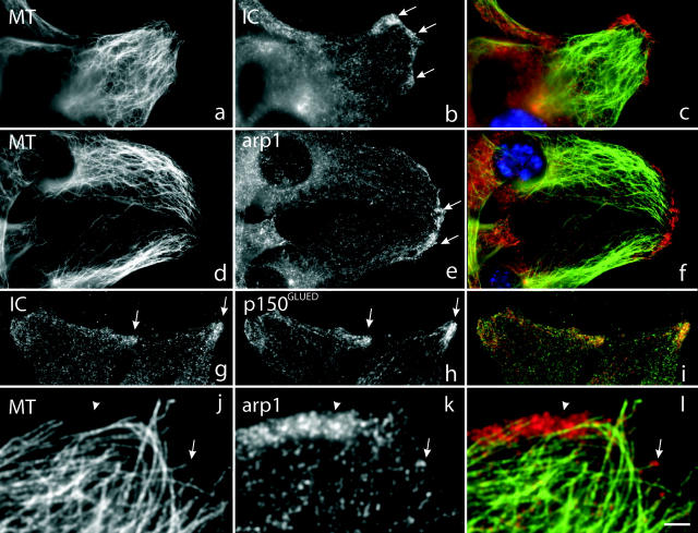
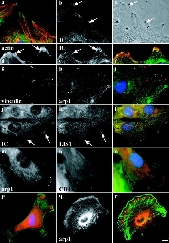
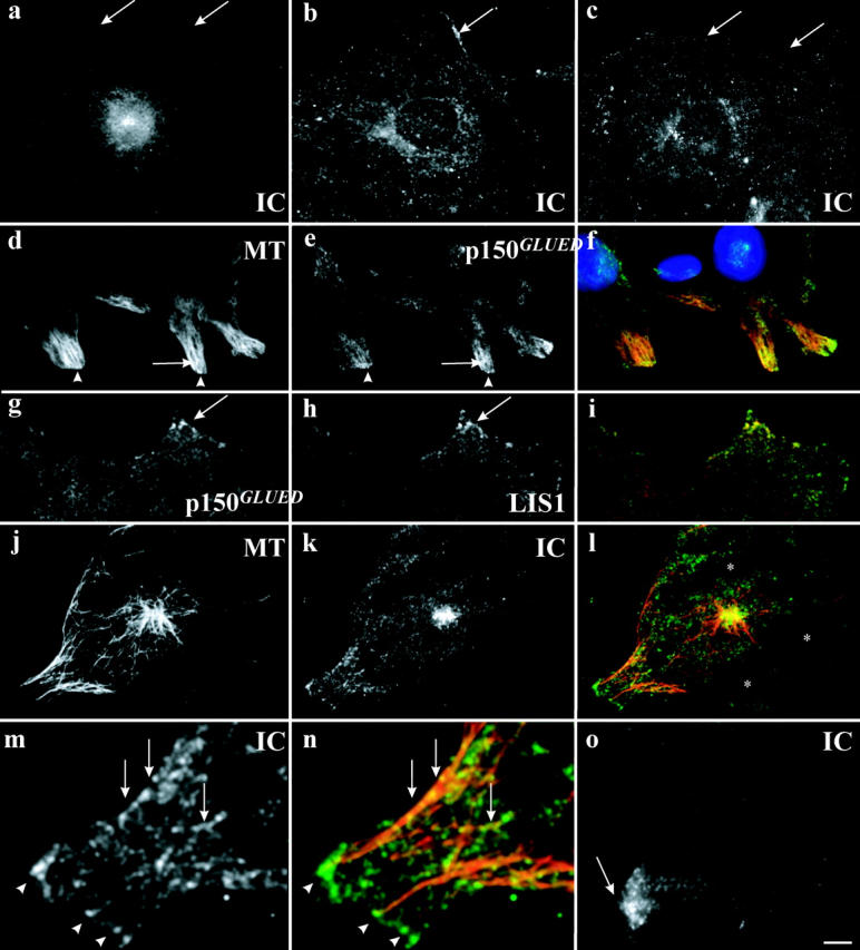
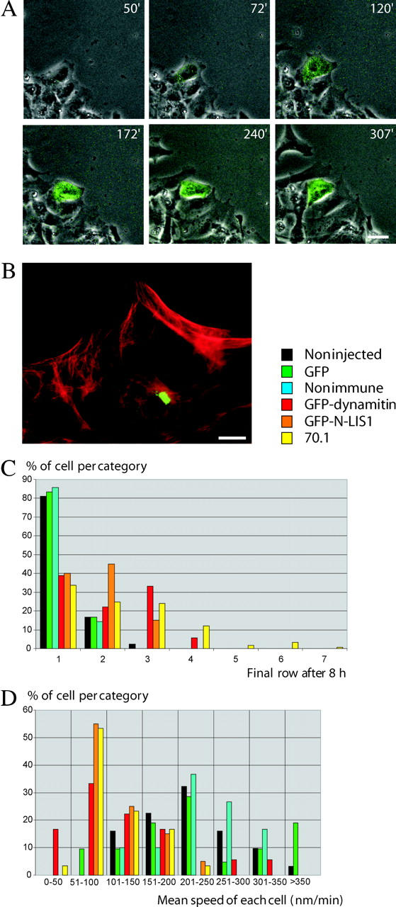
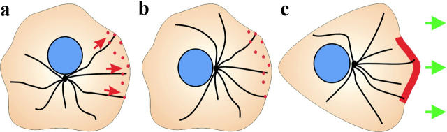
Cytoplasmic dynein has been implicated in numerous aspects of intracellular movement. We recently found dynein inhibitors to interfere with the reorientation of the microtubule cytoskeleton during healing of wounded NIH3T3 cell monolayers. We now find that dynein and its regulators dynactin and LIS1 localize to the leading cell cortex during this process. In the presence of serum, bright diffuse staining was observed in regions of active ruffling. This pattern was abolished by cytochalasin D, and was not observed in cells treated with lysophosphatidic acid, conditions which allow microtubule reorientation but not forward cell movement. Under the same conditions, using total internal reflection fluorescence microscopy, clear punctate dynein/dynactin containing structures were observed along the sides and at the tips of microtubules at the leading edge. Overexpression of dominant negative dynactin and LIS1 cDNAs or injection of antidynein antibody interfered with the rate of cell migration. Together, these results implicate a leading edge cortical pool of dynein in both early and persistent steps in directed cell movement.


Figures

Figure 1.
Figure 1.
Localization of dynein and dynactin during wound healing. Immunofluorescence microscopy of wounded NIH3T3 cell monolayers. (a–c) double labeling showing antitubulin (MT), antidynein (IC), and merged staining pattern (dynein, red); (d–f) double labeling showing antitubulin (MT) and antidynactin (arp1) and merged image (dynactin, red). (g–i) labeling showing antidynein (IC) and antidynactin (p150GLUED) and merged image (dynactin, red). (j–l) higher magnification view of double labeling showing antitubulin (MT) and antidynactin (arp1) and merged image (dynactin in red). Both diffuse (arrowheads), and punctate (arrows) staining can be observed. Cells were fixed 6 h or (g–i) 30 min after wounding. DAPI staining of nuclei is shown in blue. Bar: (a–i) 5 μm; (j–l) 2 μm.
Figure 2.
Figure 2.
Relative distribution of dynein and dynactin to other cell markers. Immunofluorescence microscopy of wounded NIH3T3 cell monolayers (a–o) and chicken embryo fibroblasts (p–r). (a–c) Enrichment of dynein IC at the leading edge (arrows) of cells at regions of lamellipodial protrusion. (a) Immunofluorescence microscopy showing antitubulin (red) and antidynein IC (green); (b) antidynein IC alone; (c) phase contrast. (d–f) Distribution of dynein (e, green in f) versus actin (d, red in f). Dynein is enriched at these sites, but not coincident with actin (p–r). (g–i) Distribution of dynactin (h, green in i) versus vinculin (g, red in i). No clear evidence for colocalization is observed. (j and k) Distribution of dynein (j, green in l) versus LIS1 (k, red in l). Colocalization at sites throughout the leading edge is observed. (m–o) Distribution of dynactin (m, red in o) versus CD44 (n, green in o). The distributions of dynactin and CD44, which is enriched in membrane ruffles (not depicted), are distinct. (p–r) Relative distribution of dynactin (q, red in p and r) versus actin (green in p and r) in low density culture of chick embryo fibroblasts. Dynectin is diffusely enriched internal to actin. Cells in a–c were fixed 8 h, in d–l 1 h, and in m–o 6 h after wounding, respectively. Bar: (a–o) 5 μm; (p–r) 10 μm.
Figure 3.
Figure 3.
Epifluorescence and TIRF microscopy during early times of wound healing. (a–c) Immunofluorescence microscopy of dynein in (a) serum-starved cells at wound edge (arrows); (b) 2 h after serum addition; (c) 2 h after LPA addition. (d–i) TIRF microscopy of serum-grown cells 1 h after wound- ing stained with (d) antitubulin; (e) antidynactin (p150GLUED); (f) merge (p150GLUED, green; DAPI staining, blue); (g) antidynactin (p150GLUED); (h) anti-LIS1; and (i) merge (LIS1, green). (j–n) TIRF microscopy of serum-starved cells exposed to LPA for 45 min stained with (j) antitubulin; (k) antidynein; (l) merge (dynein, green); (m) increased magnification of k; (n) increased magnification of l. (o) TIRF microscopy of serum-grown cells exposed to cytochalasin D for 45 min stained with antidynein. Bar: (d–f) 7 μm; (a–c, g–l, and o) 5 μm; (m and n) 2 μm.
Figure 4.
Figure 4.
Inhibition of dynein, dynactin, and LIS1 function interferes with cell migration. (A) Dual fluorescent and phase-contrast time lapse microscopy of cells wounded in the presence of serum. One cell in the field was injected with a plasmid encoding GFP-dynamitin, which was detectable as a green fluorescing signal by 60 min after injection. This cell gradually fell behind others at the wound edge. Numbers refer to minutes after wounding. (B) A monolayer of cells was wounded in the presence of serum and injected with anti-IC antibody 2 h later. After 8 h, the cells were fixed and the level of injected antibody was confirmed by immunofluorescence microscopy (antidynein antibody in green, antitubulin in red). The injected cell was seen to have fallen behind the uninjected cells at the wound edge. (C) Effects of inhibitory cDNAs and antibody on final cell position. Cell row number was determined 8 h after antibody injection or injection of inhibitory cDNA. GFP-dynamitin (red bars) and GFP-LIS1-N (orange bars) overexpressing cells as well as antidynein-injected cells (yellow bars) fell back from the leading wound edge. n = 42 (noninjected), 63 (nonimmune injected), 24 (GFP), 77 (antibody injected), 18 (GFP-dynamitin), and 20 (GFP-LIS1). (D) Distribution of speeds. The median speed was reduced by the inhibitory agents to 50–100 nm/min relative to the control values of 200–250 nm/min. Mean speeds were: GFP-dynamitin (121 ± 75 nm/min; n = 18); GFP-LIS1-N (113 ± 42 nm/min; n = 20); and antidynein injected cells (107 ± 46 nm/min; n = 30). These values were significantly lower than mean control speeds (t tests, P < 0.01): noninjected cells (227 ± 76 nm/min; n = 31); GFP overexpressors (231 ± 91 nm/min; n = 21); and nonimmune serum-injected cells (238 ± 61 nm/min; n = 30). Bars: (A) 20 μm; and (B) 10 μm.
Figure 5.
Figure 5.
Role of cytoplasmic dynein and its associated proteins during wound healing. (a and b) Cytoplasmic dynein is shown at punctate sites (red spots) at the leading cell cortex during reorientation of the microtubule network. Dynein and its associated proteins are proposed to pull on microtubules from these sites (red arrows) and/or anchor the microtubule network against retrograde forces. (c) During cell migration, dynein is enriched in regions of lamellipodial protrusion where it is proposed to regulate forward cell movement (green arrows).

References

    1. Burakov, A., E. Nadezhdina, B. Slepchenko, and V. Rodionov. 2003. Centrosome positioning in interphase cells. J. Cell Biol. 162:963–969. - PMC - PubMed
    1. Burkhardt, J.K., C.J. Echeverri, T. Nilsson, and R.B. Vallee. 1997. Overexpression of the dynamitin (p50) subunit of the dynactin complex disrupts dynein-dependent maintenance of membrane organelle distribution. J. Cell Biol. 139:469–484. - PMC - PubMed
    1. Busson, S., D. Dujardin, A. Moreau, J. Dompierre, and J.R. De Mey. 1998. Dynein and dynactin are localized to astral microtubules and at cortical sites in mitotic epithelial cells. Curr. Biol. 8:541–544. - PubMed
    1. Deacon, S.W., A.S. Serpinskaya, P.S. Vaughan, M. Lopez Fanarraga, I. Vernos, K.T. Vaughan, and V.I. Gelfand. 2003. Dynactin is required for bidirectional organelle transport. J. Cell Biol. 160:297–301. - PMC - PubMed
    1. Dohner, K., A. Wolfstein, U. Prank, C. Echeverri, D. Dujardin, R. Vallee, and B. Sodeik. 2002. Function of dynein and dynactin in herpes simplex virus capsid transport. Mol. Biol. Cell. 13:2795–2809. - PMC - PubMed

Publication types

MeSH terms