. 2003 Dec 22;163(6):1281-90.
doi: 10.1083/jcb.200307157.

Recognition of dileucine-based sorting signals from HIV-1 Nef and LIMP-II by the AP-1 gamma-sigma1 and AP-3 delta-sigma3 hemicomplexes

Affiliations

Recognition of dileucine-based sorting signals from HIV-1 Nef and LIMP-II by the AP-1 gamma-sigma1 and AP-3 delta-sigma3 hemicomplexes

Katy Janvier et al. J Cell Biol. .

Abstract

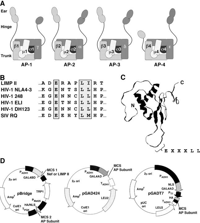
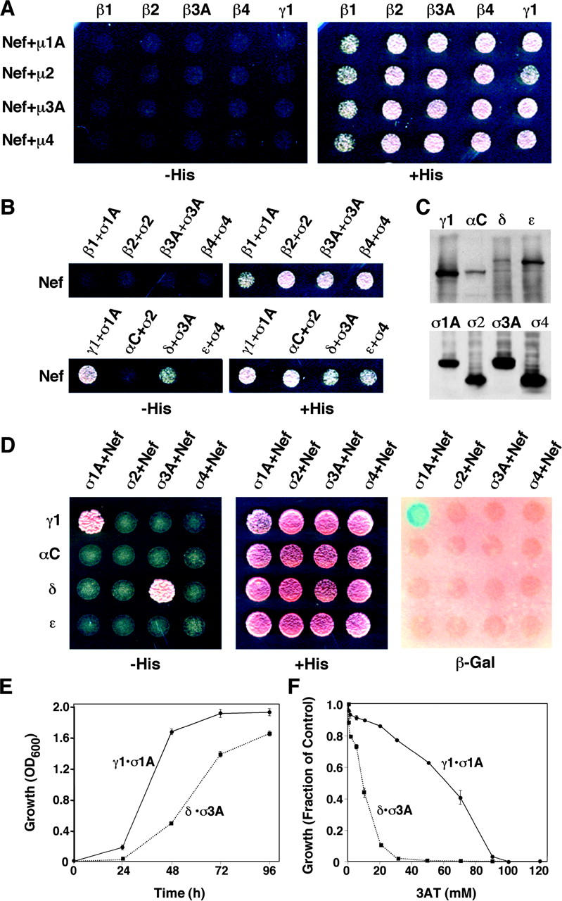
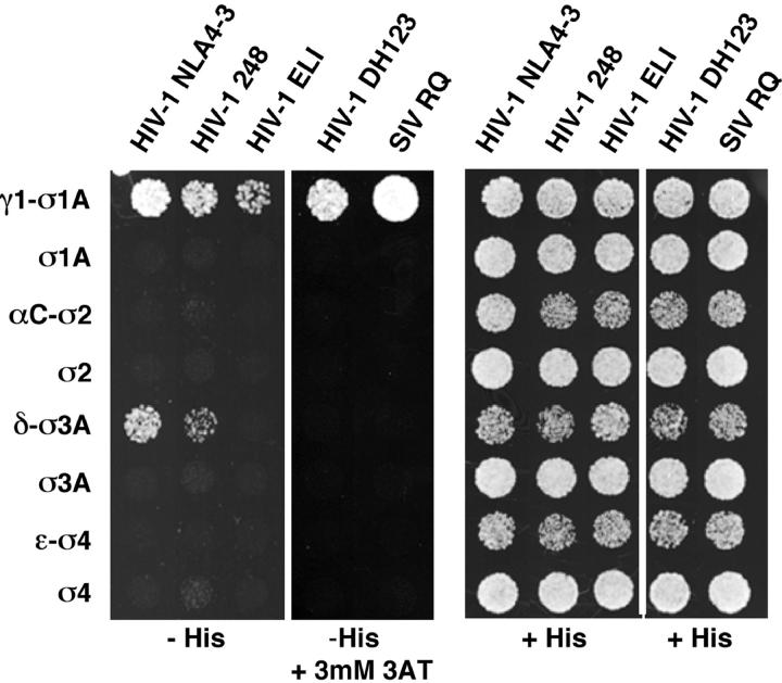
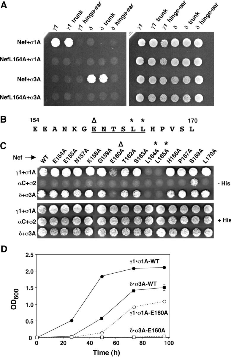
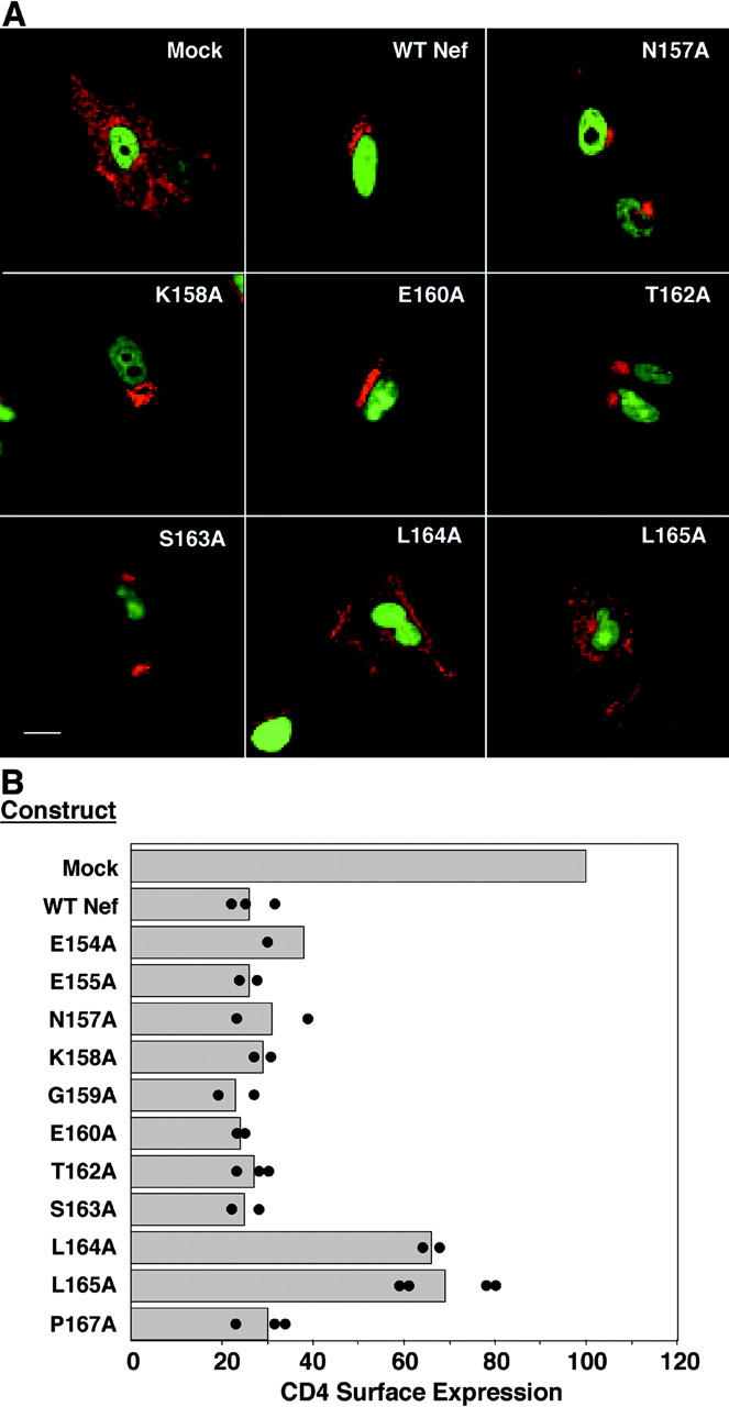
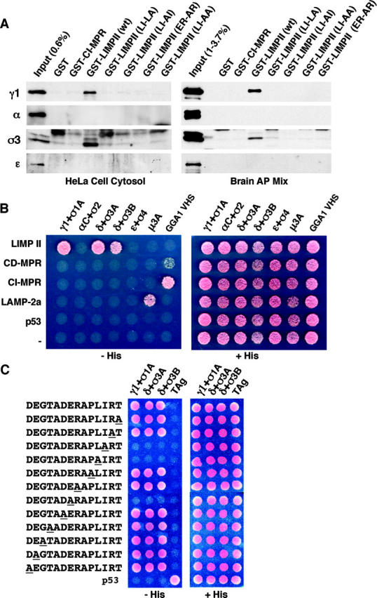
The sorting of transmembrane proteins to endosomes and lysosomes is mediated by signals present in the cytosolic tails of the proteins. A subset of these signals conform to the [DE]XXXL[LI] consensus motif and mediate sorting via interactions with heterotetrameric adaptor protein (AP) complexes. However, the identity of the AP subunits that recognize these signals remains controversial. We have used a yeast three-hybrid assay to demonstrate that [DE]XXXL[LI]-type signals from the human immunodeficiency virus negative factor protein and the lysosomal integral membrane protein II interact with combinations of the gamma and sigma1 subunits of AP-1 and the delta and sigma3 subunits of AP-3, but not the analogous combinations of AP-2 and AP-4 subunits. The sequence requirements for these interactions are similar to those for binding to the whole AP complexes in vitro and for function of the signals in vivo. These observations reveal a novel mode of recognition of sorting signals involving the gamma/delta and sigma subunits of AP-1 and AP-3.


Figures

Figure 1.
Figure 1.
Adaptors, signals, and plasmids used in this work. (A) Schematic representation of the heterotetrameric adaptor protein (AP) complexes. The designations of the generic subunits of each complex are indicated. The following subunits occur in various isoforms encoded by different genes, as indicated in parentheses: γ (γ1, γ2), μ1 (μ1A, μ1B), σ1 (σ1A, σ1B, σ1C), α (αA, αC), β3 (β3A, β3B), μ3 (μ3A, μ3B), and σ3 (σ3A, σ3B). The arrangement of the subunits was modeled after the crystal structure of the AP-2 core (Collins et al., 2002). The trunk, hinge, and ear domains of the large subunits are indicated. (B) Sequences of the dileucine-based sorting signals from LIMP-II and variants of HIV-1 and SIV Nef. The critical glutamate, leucine–leucine, leucine–isoleucine, and leucine–methionine residues are boxed. (C) NMR structure of HIV-1 Nef (BH10 isolate). The structure shown corresponds to the first of 40 structures determined by multidimensional heteronuclear NMR spectroscopy (Grzesiek et al., 1997). The locations of the NH2 and COOH termini, and of the EXXXLL sequence, are indicated. (D) Yeast expression plasmids used in two- and three-hybrid assays. The pBridge plasmid expresses two proteins, GAL4BD fused to Nef or the LIMP-II cytosolic tail (multiple cloning site 1, MCS 1) and an AP subunit (MCS 2). The pGAD424 and pGADT7 plasmids drive high-level expression of GAL4AD fused to another AP subunit.
Figure 2.
Figure 2.
Three-hybrid analysis of the interaction of HIV-1 Nef (NLA4-3 variant) with different AP subunits. (A) GAL4BD-Nef and the μ subunits were expressed from pBridge, whereas the β subunits and γ1 were expressed as fusions with GAL4AD from pGADT7. (B) GAL4BD-Nef and the σ subunits were expressed from pBridge, whereas the β1, β2, β3A, β4, γ1, αC, δ, and ɛ were expressed as fusions with GAL4AD from pGADT7. (C) Immunoblot analysis (using anti-HA) of AP subunits in yeast cells cotransformed with γ1–σ1A, αC–σ2, δ–σ3A, or ɛ–σ4. The subunits were expressed as in B, and all subunits were tagged with the HA epitope. (D) GAL4BD-Nef and the σ subunits were expressed from pBridge, whereas γ1, αC, δ, and ɛ were expressed as fusions with GAL4AD from pGADT7. In A, B, and D, interactions were evidenced by growth on agar plates made with medium without histidine (−His) or by expression of β-galactosidase (β-Gal) activity. (E) Time course of growth of yeast strains coexpressing Nef with either γ1–σ1A (γ1•σ1A) or δ–σ3A (δ•σ3A) was measured by optical density at 600 nm (OD600). (F) Inhibition by 3AT of growth of yeast strains coexpressing Nef with either γ1–σ1A (γ1•σ1A) or δ–σ3A (δ•σ3A). Growth was measured by optical density at 600 nm after 48 h in culture. The value 1.0 on the y axis corresponds to the growth of each strain in the absence of 3AT. The expression of Nef and AP subunits from plasmids was as described in D. Values are the mean ± SD of triplicate determinations.
Figure 3.
Figure 3.
Interaction of naturally occurring Nef variants with AP subunits. Three-hybrid analysis of the interaction of Nef from different isolates of HIV-1 or SIV fused to GAL4BD (pBridge) with different σ subunits (pBridge) and γ1, αC, δ, and ɛ subunits fused to GAL4AD (pGADT7). The sequences of the dileucine-based sorting signals from each of these constructs are indicated in Fig. 1 B. Interactions were evidenced by growth on agar plates made with medium without histidine (−His). The HIV-1 DH123 and SIV RQ Nef variants self-activated in this assay and had to be tested for growth in the presence of 3 mM 3AT.
Figure 4.
Figure 4.
Mutational analyses of AP subunits and HIV-1 Nef. (A) Three-hybrid analysis of the interaction of Nef (NLA4-3 variant) or a NefL164A mutant GAL4BD (pBridge) with σ1A or σ3A (pBridge) and γ1, δ, or the trunk or hinge–ear fragments from these subunits fused to GAL4AD (pGADT7). (B) Sequence of the dileucine-based sorting signal (underlined) and flanking residues (154–170) in the NLA4-3 variant of HIV-1 Nef. In B and C, the critical glutamate and leucine residues are indicated by an open triangle and asterisks, respectively. (C) Three-hybrid analysis of the interaction of full-length, wild-type (WT) Nef (NLA4-3 variant) or alanine mutants of this Nef (pBridge) with σ1A, σ2, or σ3A (pBridge) and γ1, αC, or δ (pGADT7), as indicated in the figure. Interactions were detected by growth on agar plates made with medium without histidine (−His). (D) Time course of growth of yeast strains coexpressing wild-type (WT) or the E160A Nef mutant with either γ1–σ1A (γ1•σ1A) or δ–σ3A (δ•σ3A). Plasmid constructs were as described in C. Growth was measured by changes in the optical density at 600 nm (OD600). Values are the mean ± SD of triplicate determinations.
Figure 5.
Figure 5.
Down-regulation of CD4 by Nef or Nef mutants. (A) Analysis by immunofluorescence microscopy. HeLa cells were cotransfected with pCMV-CD4, pEGFP encoding GFP-histone H2B, and pCI-neo encoding either wild-type Nef (NLA4-3 variant) or Nef mutants. At 24 h after transfection, cells were fixed, permeabilized, and immunostained for CD4 (red). Cotransfected cells were identified by the expression of GFP-histone H2B in the nucleus (green). Images corresponding to selected Nef constructs are shown. Bar, 25 μm. (B) Analysis by FACS®. HeLa cells were cotransfected with pCMV-CD4, pCMV-CD8, and pCI-neo encoding either wild-type Nef (NLA4-3 variant) or Nef mutants. At 24 h after transfection, cells were coimmunostained with allophycocyanin-conjugated anti-CD4 and phycoerythrin-conjugated anti-CD8 antibodies. The percentage of CD8+ cells that also expressed CD4 on the surface was quantified by FACS® analysis, as described in Materials and methods. The results obtained in different determinations for each Nef construct are indicated by the individual dots. Bars represent the mean values for each Nef construct.
Figure 6.
Figure 6.
Interactions of the dileucine-based sorting signal from LIMP-II with AP complexes. (A) GST pull-down assays. GST fused to the cytosolic tail of the CI-MPR, LIMP-II, or mutants of this latter construct were incubated with either a cytosolic extract prepared from HeLa cells or a mixture of AP complexes prepared from bovine brain clathrin-coated vesicles. The LIMP-II tail mutants contain the amino acid substitutions indicated in the figure relative to the LIMP-II sequence shown in Fig. 1 B. Bound AP complexes were revealed by immunoblot analysis with antibodies specific for each complex. Some of the AP subunits (most notably σ3 and α in bovine brain) appeared as doublets. (B) Three-hybrid analysis of the interaction of the LIMP-II, CD-MPR, CI-MPR, and LAMP-2a cytosolic tails fused to GAL4BD (pBridge) with γ1, αC, δ, and ɛ (pBridge) and different σ subunits; μ3A and the VHS domain of GGA1 fused to GAL4AD (pGAD424). Interactions were detected by growth in the absence of histidine (−His). (C) Alanine-scan mutagenesis analysis of the interaction of LIMP-II cytosolic tail mutants with γ1–σ1A (γ1 + σ1A), δ–σ3A (δ + σ3A), or δ–σ3B (δ + σ3B) using the yeast-three hybrid system. Mutated residues are underlined. Plasmid constructs were as specified in B. The SV40 large T antigen (TAg) and p53 were used as controls. Interactions were detected by growth in the absence of histidine (−His).

References

    1. Aguilar, R.C., H. Ohno, K.C. Roche, and J.S. Bonifacino. 1997. Functional domain mapping of the clathrin-associated adaptor medium chains mu1 and mu2. J. Biol. Chem. 272:2760–2766. - PubMed
    1. Aiken, C., J. Konner, N.R. Landau, M.E. Lenburg, and D. Trono. 1994. Nef induces CD4 endocytosis: requirement for a critical dileucine motif in the membrane-proximal CD4 cytoplasmic domain. Cell. 76:853–864. - PubMed
    1. Barriocanal, J.G., J.S. Bonifacino, L. Yuan, and I.V. Sandoval. 1986. Biosynthesis, glycosylation, movement through the Golgi system, and transport to lysosomes by an N-linked carbohydrate-independent mechanism of three lysosomal integral membrane proteins. J. Biol. Chem. 261:16755–16763. - PubMed
    1. Boehm, M., R.C. Aguilar, and J.S. Bonifacino. 2001. Functional and physical interactions of the adaptor protein complex AP-4 with ADP-ribosylation factors (ARFs). EMBO J. 20:6265–6276. - PMC - PubMed
    1. Bonifacino, J.S., and L.M. Traub. 2003. Signals for sorting of transmembrane proteins to endosomes and lysosomes. Annu. Rev. Biochem. 72:395–447. - PubMed

MeSH terms