. 2004 Jan 6;101(1):378-83.
doi: 10.1073/pnas.0304699101. Epub 2003 Dec 22.

Heptahelical domain of metabotropic glutamate receptor 5 behaves like rhodopsin-like receptors

Affiliations

Heptahelical domain of metabotropic glutamate receptor 5 behaves like rhodopsin-like receptors

Cyril Goudet et al. Proc Natl Acad Sci U S A. .

Abstract

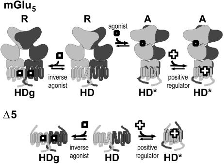
Although agonists bind directly in the heptahelical domain (HD) of most class-I rhodopsin-like G protein coupled receptors (GPCRs), class-III agonists bind in the extracellular domain of their receptors. Indeed, the latter possess a large extracellular domain composed of a cysteine-rich domain and a Venus flytrap module. Both the low sequence homology and the structural organization of class-III GPCRs raised the question of whether or not the HD of these receptors functions the same way as rhodopsin-like GPCRs. Here, we show that the HD of metabotropic glutamate receptor 5 (mGlu(5)) displays the same agonist-independent constitutive activity as the wild-type receptor. Moreover, we show that the noncompetitive antagonist MPEP [2-methyl-6-(phenylethynyl)-pyridine hydrochloride] and the positive allosteric modulator DFB (3,3'-difluorobenzaldazine) act as inverse agonist and full agonist, respectively, on the mGlu(5) HD in the absence of the extracellular domain. This finding illustrates that, like rhodopsin-like receptors, the HD of mGluRs can constitutively couple to G proteins and be negatively and positively regulated by ligands. These data show that the HD of mGluRs behave like any other class-I GPCRs in terms of G protein coupling and regulation by various types of ligands.


Figures

Fig. 1.
Fig. 1.
Cell surface expression of mGlu5, Δ5, and Δ5Δ. (A) Schematic representation of mGlu5, Δ5, and Δ5Δ and location of the sites of truncation. The open box represents the HA tag, and the gray box corresponds to the signal peptide of mGlu5.(B) Surface expression of mGlu5, Δ5, and Δ5Δ in HEK 293 cells was detected by immunofluorescence on nonpermeabilized cells. (C) Quantification of cell surface expression of mGlu5, Δ5, and Δ5Δ by ELISA on intact cells. Cells were transfected with 0.6, 5, and 5 μg of plasmids expressing mGlu5, Δ5, and Δ5Δ, respectively.
Fig. 2.
Fig. 2.
Like mGlu5, Δ5 and Δ5Δ are constitutively active. (A) IP production measured in mGlu5, Δ5, and Δ5Δ or mock-transfected HEK 293 cells under basal conditions (open bars) or in the presence of 1 mM Glu (filled bars). Basal IP formation in mock-transfected cell is highlighted by a dotted line. Data correspond to the ratio between total IP produced by the cells and the total radioactivity remaining in the membranes plus the produced IPs. (B) MPEP decreased the basal IP production in HEK293 cells expressing mGlu5 (open circles) and Δ5Δ receptors (filled circles). Results are expressed as the percentage of the basal IP production measured in the absence of MPEP.
Fig. 3.
Fig. 3.
DFB potentiates agonist-induced activity of wild-type mGlu5. (A) Effect of increasing concentrations of glutamate in the absence (CTR, open circles) or presence (DFB, filled circles) of 100 μM DFB on IP production in cells expressing mGlu5.(B) Effect of increasing concentration of DFB was measured on cells expressing mGlu5 in the absence (open circles) or presence of 10 nM quisqualate (filled circles). (Inset) The effect of 10 nM quisqualate on intracellular Ca2+ concentration was measured under control condition (C) or in the presence of 100 μM DFB (DFB). Vertical bar represents a change in the fluorescence signal of 1,000 units. Data are expressed as the percentage of the maximal effect measured with quisqualate plus DFB.
Fig. 4.
Fig. 4.
Direct activation of Δ5Δ by the positive allosteric regulator DFB. (A) Effect of increasing doses of DFB on Δ5Δ. DFB dose-dependently activates the truncated receptor Δ5Δ. The curve has been normalized such that the basal response is zero and the maximum is 100%. (Inset) IP formation (% above the basal) was induced by DFB only in cells expressing Δ5Δ and not in cells expressing mGlu5. (B) Direct stimulation of Δ5 and Δ5Δ by 100 μM DFB as revealed by intracellular Ca2+ measurement with Fluo-4. (C) Activity of mGlu5 (squares) and Δ5Δ (circles) as a function of their membrane expression. Cells were transfected with increasing amounts of cDNA coding for these receptors, and surface expression of mGlu5 and Δ5Δ was measured by ELISA on intact cells. Basal (open symbols) and glutamate-(1 mM) or DFB-(1 mM) (filled symbols) induced IP formation was measured in parallel.
Fig. 5.
Fig. 5.
MPEP inhibits partially DFB-induced activity on Δ5Δ. (A) Effect of 10, 30, and 100 nM MPEP on IP production induced by 300 μM DFB in cells expressing Δ5Δ. (B) Effect of 10 and 100 nM MPEP on intracellular Ca2+ release induced by 300 μM DFB on cells expressing Δ5Δ.
Fig. 6.
Fig. 6.
Schematic representation of the possible action of inverse agonists and positive modulators of mGlu5. (Upper) The constitutive dimer of mGlu5 is shown to be composed of a VFTM (top), a CRD (middle), and a HD. The HD is proposed to oscillate between a slightly active state (HD) and a totally inactive ground state (HDg), the latter being stabilized by inverse agonist. This equilibrium can occur even though the dimer of VFTM stays in the resting state (R). The dimer of VFTMs is assumed to reach an active orientation (A) in the presence of agonist, leading to the stabilization of a fully active state of the dimer of HDs (HD*). The positive allosteric modulator, DFB, is proposed to bind with a higher affinity on HD*, stabilizing the fully active state of the receptor, leading to an increased affinity of the receptor for agonists (19). (Lower)Inthe absence of the large extracellular domain, the HD can reach more freely the fully active state HD*, allowing the positive modulators to act as full agonists.

References

    1. Bockaert, J. & Pin, J.-P. (1999) EMBO J. 18, 1723-1729. - PMC - PubMed
    1. Bockaert, J., Claeysen, S., Becamel, C., Pinloche, S. & Dumuis, A. (2002) Int. Rev. Cytol. 212, 63-132. - PubMed
    1. Fredriksson, R., Lagerstrom, M. C., Lundin, L. G. & Schioth, H. B. (2003) Mol. Pharmacol. 63, 1256-1272. - PubMed
    1. Lefkowitz, R. J., Cotecchia, S., Samama, P. & Costa, T. (1993) Trends Pharmacol. Sci. 14, 303-307. - PubMed
    1. Leff, P. (1995) Trends Pharmacol. Sci. 16, 89-97. - PubMed

Publication types

MeSH terms