. 2003 Dec;1(3):E72.
doi: 10.1371/journal.pbio.0000072. Epub 2003 Dec 22.

Architecture and selectivity in aquaporins: 2.5 a X-ray structure of aquaporin Z

Affiliations

Architecture and selectivity in aquaporins: 2.5 a X-ray structure of aquaporin Z

David F Savage et al. PLoS Biol. 2003 Dec.

Abstract

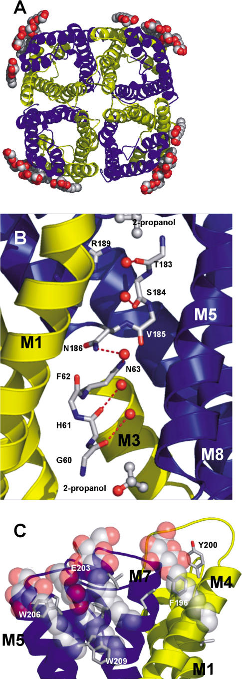
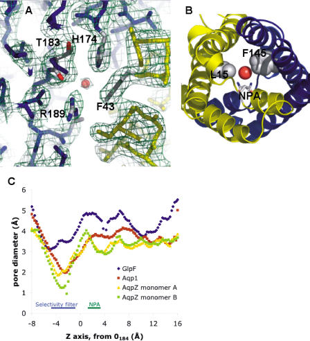
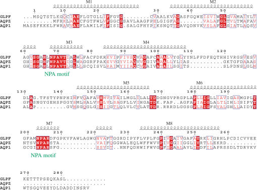
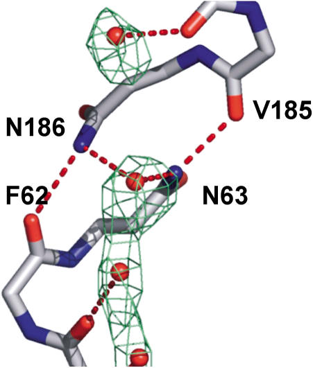
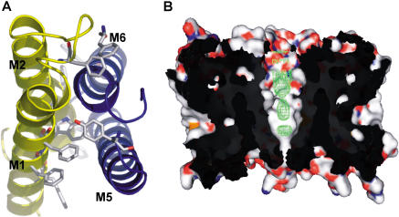
Aquaporins are a family of water and small molecule channels found in organisms ranging from bacteria to animals. One of these channels, the E. coli protein aquaporin Z (AqpZ), has been shown to selectively conduct only water at high rates. We have expressed, purified, crystallized, and solved the X-ray structure of AqpZ. The 2.5 A resolution structure of AqpZ suggests aquaporin selectivity results both from a steric mechanism due to pore size and from specific amino acid substitutions that regulate the preference for a hydrophobic or hydrophilic substrate. This structure provides direct evidence on the molecular mechanisms of specificity between water and glycerol in this family of channels from a single species. It is to our knowledge the first atomic resolution structure of a recombinant aquaporin and so provides a platform for combined genetic, mutational, functional, and structural determinations of the mechanisms of aquaporins and, more generally, the assembly of multimeric membrane proteins.


Conflict of interest statement

The authors have declared that no conflicts of interest exist.

Figures

Figure 1
Figure 1. Structure of AqpZ
Three-dimensional fold of AqpZ with the quasi-2-fold related segments in yellow (residues 1–117) and blue (residues 188–231). (A) Cartoon representation of the AqpZ tetramer with OG detergent molecules represented as spheres; view is from the periplasmic side. Atoms are colored according to atom type (red, oxygen; gray, carbon; blue, nitrogen; yellow, sulfur). (B) Cartoon representation of the AqpZ monomer, with M2 and M6 removed for ease of viewing. Single-file water is shown hydrogen-bonding to carbonyls of main chain. Central water is shown accepting a hydrogen bond from the NH2 group of Asn63 and Asn186. Sidechains of the selectivity filter are also shown. Isopropanol molecules located in density are shown as sticks, just outside the channel. (C) A view from the membrane plane of the OG micelle interactions with the periplasmic segment of AqpZ. OG molecules pack against the aromatic sidechains, while making hydrogen bonds with the main chain carbonyls, E203, and each other. All figures were made with PyMOL (DeLano 2003).
Figure 2
Figure 2. Channel Constriction in Aquaporins
(A) A view of the aquaporin selectivity filter from the periplasmic side. Experimental electron density (2Fobs – Fcalc ) is contoured at 1.1 σ. (B) Secondary constriction at the NPA motif due to F145 and L15. The drawn water is HOH1032, hydrogen-bonded to the NPA motif asparagines. (C) Pore diameters for the aquaporin X-ray structures, calculated with HOLE2. The AqpZ monomers (protomers) A and B refer to the crystallographically distinct monomers in the unit cell.
Figure 3
Figure 3. Sequence Alignment of Aquaporins with Known Structure
Alignment of GlpF, AqpZ, and AQP1 is numbered according to GlpF. Helices are shown and labeled M1–M8. Residue positions of similar chemical nature are shown in blue boxes; identical residues are shown in red.
Figure 4
Figure 4. Water at the NPA Region
N63 and N186 donate hydrogen bonds to the central water by projecting their NH2 moieties into the pore. This conformation is aided by a hydrogen bond from the adjacent carbonyls of V185 and F62, respectively. Experimental electron density (2Fobs – Fcalc) is contoured at 0.7 σ.
Figure 5
Figure 5. Oligomerization of AqpZ
(A) Protomer–protomer interface. The interface is composed of helices M1 and M2 from one protomer (yellow) and M5 and M6 from a second protomer (blue). The helices participate in knobs into holes packing, and the interface is extensive (3,340 Δ2) owing to the large number of aromatic residues. (B) Abstruse electron density along the 4-fold axis (colored in green). Two protomers are display in surface rendering. The experimental electron density (2Fobs – Fcalc) is contoured at 1.1 σ.

References

    1. Abramson J, Smirnova I, Kasho V, Verner G, Kaback HR, et al. Structure and mechanism of the lactose permease of Escherichia coli. Science. 2003;301:610–615. - PubMed
    1. Bernal JD, Fowler RH. A theory of water and ionic solution, with particular reference to hydrogen and hydroxyl ions. J Chem Phys. 1933;1:515–548.
    1. Borgnia MJ, Agre P. Reconstitution and functional comparison of purified GlpF and AqpZ, the glycerol and water channels from Escherichia coli. Proc Natl Acad Sci U S A. 2001;98:2888–2893. - PMC - PubMed
    1. Borgnia MJ, Kozono D, Calamita G, Maloney PC, Agre P. Functional reconstitution and characterization of AqpZ, the E. coli water channel protein. J Mol Biol. 1999a;291:1169–1179. - PubMed
    1. Borgnia M, Nielsen S, Engel A, Agre P. Cellular and molecular biology of the aquaporin water channels. Annu Rev Biochem. 1999b;68:425–458. - PubMed

Publication types

MeSH terms

Associated data