. 2004 Jan;78(2):669-82.
doi: 10.1128/jvi.78.2.669-682.2004.

Characterization of the RNA components of a putative molecular switch in the 3' untranslated region of the murine coronavirus genome

Affiliations

Characterization of the RNA components of a putative molecular switch in the 3' untranslated region of the murine coronavirus genome

Scott J Goebel et al. J Virol. 2004 Jan.

Abstract

RNA virus genomes contain cis-acting sequence and structural elements that participate in viral replication. We previously identified a bulged stem-loop secondary structure at the upstream end of the 3' untranslated region (3' UTR) of the genome of the coronavirus mouse hepatitis virus (MHV). This element, beginning immediately downstream of the nucleocapsid gene stop codon, was shown to be essential for virus replication. Other investigators discovered an adjacent downstream pseudoknot in the 3' UTR of the closely related bovine coronavirus (BCoV). This pseudoknot was also shown to be essential for replication, and it has a conserved counterpart in every group 1 and group 2 coronavirus. In MHV and BCoV, the bulged stem-loop and pseudoknot are, in part, mutually exclusive, because of the overlap of the last segment of the stem-loop and stem 1 of the pseudoknot. This led us to hypothesize that they form a molecular switch, possibly regulating a transition occurring during viral RNA synthesis. We have now performed an extensive genetic analysis of the two components of this proposed switch. Our results define essential and nonessential components of these structures and establish the limits to which essential parts of each element can be destabilized prior to loss of function. Most notably, we have confirmed the interrelationship of the two putative switch elements. Additionally, we have identified a pseudoknot loop insertion mutation that appears to point to a genetic interaction between the pseudoknot and a distant region of the genome.


Figures
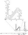
FIG. 1.
FIG. 1.
Landscape of the MHV 3′ UTR. The organization of the 31.3-kb MHV genome is shown at the bottom. Above this is an expanded view of the secondary structure of the 301-nt 3′ UTR, which includes the bulged stem-loop demonstrated previously for nt 234 through 301 (6, 7), a pseudoknot comprising nt 185 through 238 (37), and a complex structure containing multiple stem-loops and bulges proposed for nt 1 through 156 (18). At the top is a detailed view of the upstream end of the 3′ UTR. Stem segments B, C, D, and F are shown as determined by Hsue et al. (6) (reprinted from reference 6). Pseudoknot stems S1 and S2 and loops L1 and L2 are labeled as determined by Williams et al. (37). Nucleotides are numbered from the first base at the 3′ end of the genome, excluding poly(A), and the N-gene stop codon is boxed. Broken lines in the upper diagram indicate alternative base pairings for stem segment F or pseudoknot stem 1.
FIG. 2.
FIG. 2.
Selection of MHV 3′ UTR mutants by targeted RNA recombination. Interspecies chimeric viruses fMHV (12) and fMHV.v2 each contain the ectodomain-encoding region of the FIPV S gene (hatched rectangle) and are consequently able to grow in feline cells, but not in murine cells. Mutated 3′ UTRs (black rectangles) can be transduced into either one of these recipient viral genomes by recombination with donor RNA transcribed from derivatives of plasmid pMH54. A single crossover (solid line) within the HE gene should generate a recombinant that has simultaneously reacquired the MHV S ectodomain and the ability to grow in murine cells and has also incorporated the mutated 3′ UTR. A potential second crossover (broken line), downstream of the S-gene ectodomain region, would produce a recombinant retaining the wild-type 3′ UTR. Bracketed horizontal bars indicate the boundaries of the region in which each type of crossover could occur. For pMH54-derived RNA, [1] and [HE] indicate 467- and 1,185-nt fragments of the 5′ end of the MHV genome and HE gene, respectively, as described previously (12). In the fMHV.v2 genome, [M] and [4] indicate 126- and 169-nt fragments of the M gene and gene 4, respectively, as described previously for plasmid pA122 (3).
FIG. 3.
FIG. 3.
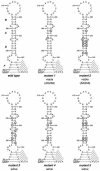
Stabilization or destabilization of the bulged stem-loop. For each mutant, nucleotides that were changed from those of the wild-type sequence are circled. Nucleotides are numbered from the first base at the 3′ end of the genome, excluding poly(A). Broken lines indicate alternative base pairings for stem segment F or pseudoknot stem 1. Unless indicated otherwise, viable mutants have a wild-type phenotype.
FIG. 4.
FIG. 4.
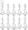
Truncation and more localized destabilization of the bulged stem-loop. For each mutant, nucleotides that were changed from those of the wild-type sequence are circled. Nucleotides are numbered from the first base at the 3′ end of the genome, excluding poly(A), and the N-gene stop codon is boxed. Broken lines indicate alternative base pairings for stem segment F or pseudoknot stem 1. The black arrowhead indicates a deletion. Unless indicated otherwise, viable mutants have a wild-type phenotype.
FIG. 5.
FIG. 5.
Alteration of the bulge bases between stem segments C and D. For each mutant, nucleotides that were changed from those of the wild-type sequence are circled. Nucleotides are numbered from the first base at the 3′ end of the genome, excluding poly(A), and the N-gene stop codon is boxed. Broken lines indicate alternative base pairings for stem segment F or pseudoknot stem 1.
FIG. 6.
FIG. 6.
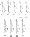
Disruption or destabilization of pseudoknot stem 2. To clarify the relationships among the components of the pseudoknot, we show a more standard representation of pseudoknot stem 1 here (different from the schematic in Fig. 1). For each mutant, nucleotides that were changed from those of the wild-type sequence are circled. Nucleotides are numbered from the first base at the 3′ end of the genome, excluding poly(A). Broken lines indicate alternative base pairings for stem segment F or pseudoknot stem 1. Unless indicated otherwise, viable mutants have a wild-type phenotype.
FIG. 7.
FIG. 7.
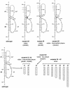
Destabilization of pseudoknot components. For each mutant, nucleotides that were changed from those of the wild-type sequence are circled. Nucleotides are numbered from the first base at the 3′ end of the genome, excluding poly(A). Broken lines indicate alternative base pairings for stem segment F or pseudoknot stem 1. The black arrowhead (mutant 28) indicates a deletion. The white arrowhead (mutants 31 through 40) indicates an insertion of variable size. Unless otherwise indicated, viable mutants have a wild-type phenotype.
FIG. 8.
FIG. 8.
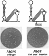
Plaque phenotype of loop 1 insertion mutant Alb391 compared to wild type. Plaque titrations were performed on mouse L2 cells at 37°C. Monolayers were stained with neutral red at 48 h postinfection and photographed 24 h later.
FIG. 9.
FIG. 9.
Essential nature of the overlap of stem segment F and pseudoknot stem 1. Mutants 41 through 43 have been reported previously (designated mutants MFL, MFR, and MFLR, respectively) (6). For each mutant, nucleotides that were changed from those of the wild-type sequence are circled. Nucleotides are numbered from the first base at the 3′ end of the genome, excluding poly(A), and the N-gene stop codon is boxed. Broken lines indicate alternative base pairings for stem segment F or pseudoknot stem 1. Unless indicated otherwise, viable mutants have a wild-type phenotype.
FIG. 10.
FIG. 10.
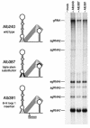
RNA synthesis by 3′ UTR mutants Alb387 (mutant 45 in Fig. 9) and Alb391 (mutant 33 in Fig. 7) compared to wild-type Alb240. Infected or mock-infected cells were metabolically labeled with 33Pi, and RNA was isolated and analyzed as described in Materials and Methods. gRNA, genomic RNA.
FIG. 11.
FIG. 11.
Conservation of the 3′ UTR bulged stem-loop and overlapping pseudoknot structure between group 2 coronaviruses (exemplified by MHV) and the SARS-associated coronavirus (SARS-CoV).

Similar articles

Cited by

References

    1. Baric, R. S., and B. Yount. 2000. Subgenomic negative-strand RNA function during mouse hepatitis virus infection. J. Virol. 74:4039-4046. - PMC - PubMed
    1. Dalton, K., R. Casais, K. Shaw, K. Stirrups, S. Evans, P. Britton, T. D. K. Brown, and D. Cavanagh. 2001. cis-acting sequences required for coronavirus infectious bronchitis virus defective-RNA replication and packaging. J. Virol. 75:125-133. - PMC - PubMed
    1. de Haan, C. A. M., H. Volders, C. A. Koetzner, P. S. Masters, and P. J. M. Rottier. 2002. Coronaviruses maintain viability despite dramatic rearrangements of the strictly conserved genome organization. J. Virol. 76:12491-12493. - PMC - PubMed
    1. Giedroc, D. P., C. A. Theimer, and P. L. Nixon. 2000. Structure, stability, and function of RNA pseudoknots involved in stimulating ribosomal frameshifting. J. Mol. Biol. 298:167-185. - PMC - PubMed
    1. Horton, R. M., and L. R. Pease. 1991. Recombination and mutagenesis of DNA sequences using PCR, p. 217-247. In M. J. McPherson (ed.), Directed mutagenesis, a practical approach. IRL Press, New York, N.Y.

Publication types

LinkOut - more resources