. 2004 Jan;78(2):821-33.
doi: 10.1128/jvi.78.2.821-833.2004.

Functional analysis of the human papillomavirus type 16 E1=E4 protein provides a mechanism for in vivo and in vitro keratin filament reorganization

Affiliations

Functional analysis of the human papillomavirus type 16 E1=E4 protein provides a mechanism for in vivo and in vitro keratin filament reorganization

Qian Wang et al. J Virol. 2004 Jan.

Abstract

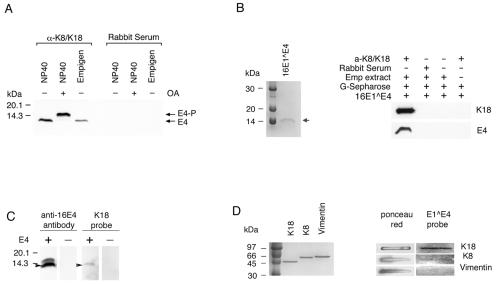
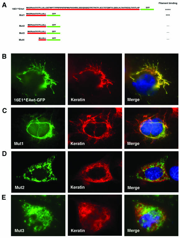
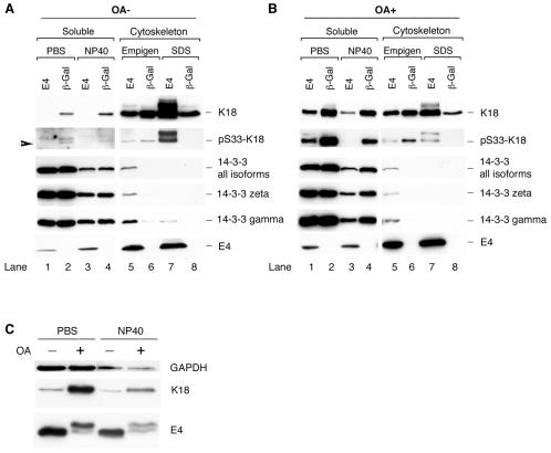
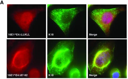
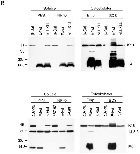
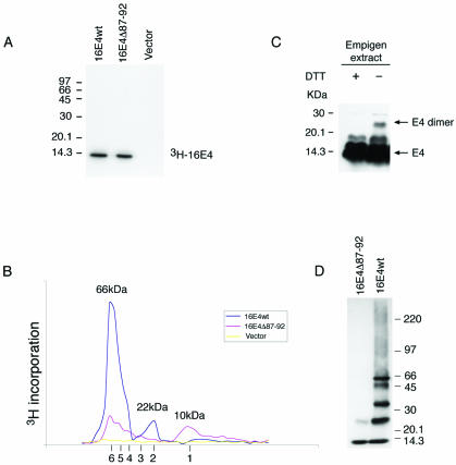
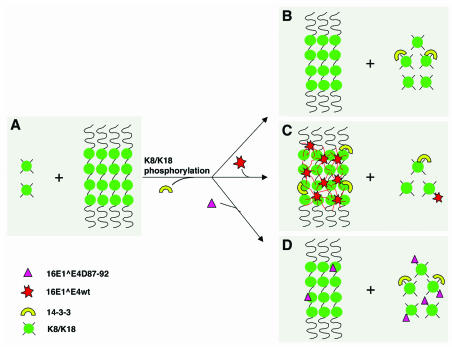
High-risk human papillomaviruses, such as human papillomavirus type 16 (HPV16), are the primary cause of cervical cancer. The HPV16 E1=E4 protein associates with keratin intermediate filaments and causes network collapse when expressed in epithelial cells in vitro. Here, we show that keratin association and network reorganization also occur in vivo in low-grade cervical neoplasia caused by HPV16. The 16E1=E4 protein binds to keratins directly and interacts strongly with keratin 18, a member of the type I intermediate-filament family. By contrast, 16E1=E4 bound only weakly to keratin 8, a type II intermediate-filament protein, and showed no detectable affinity for the type III protein, vimentin. The N-terminal 16 amino acids of the 16E1=E4 protein, which contains the YPLLXLL motif that is conserved among supergroup A viruses, were sufficient to target green fluorescent protein to the keratin network. When expressed in the SiHa cervical epithelial cell line, the full-length 16E1=E4 protein caused an almost total inhibition of keratin dynamics, despite the phosphorylation of keratin 18 at serine 33, which normally leads to 14-3-3-mediated keratin solubilization. Mutant 16E1=E4 proteins which lack the LLKLL motif, or which have lost amino acids from their C termini, and which were compromised in the ability to associate with keratins did not disturb normal keratin dynamics. 16E1=E4 was found to exist as dimers and hexamers, whereas a C-terminal deletion mutant (16E1=E4Delta87-92) existed as monomers and formed multimeric structures only poorly. Considered together, our results suggest that by associating with keratins through its N terminus, and by associating with itself through its C terminus, 16E1=E4 may act as a keratin cross-linker and prevent the movement of keratins between the soluble and insoluble compartments. The increase in avidity associated with multimeric binding may contribute to the ability of 16E1=E4 to sequester its cellular targets in the cytoplasm.


Figures

FIG. 1.
FIG. 1.
HPV16E1E4 is associated with reorganized keratin network in vivo and in vitro. (A and D) Tissue sections from cervical lesions caused by HPV16 were triple stained for 16E1E4 (green), K13 (red), and DNA (blue). The dotted lines indicate the position of the basal layer. The images were taken using a 10× objective. The cells enclosed within boxes are shown at higher magnification in panels B, C, and E (images captured using a 100× objective). (B) Colocalization of 16E1E4 and K13 filaments. The image corresponds to the right-hand box in panel A. (C) Association of reorganized keratin networks with 16E1E4. The image corresponds to the left-hand box in panel A. (E) Association of reorganized keratin networks with 16E1E4. The image corresponds to the left-hand box in panel D. The small box in the top right-hand corner of panel D, which contains cells with normal keratin staining, is shown enlarged in the inset. (F) 16E1E4-associated keratin network collapse in cultured epithelial cells is shown for comparison 24 h after infection of SiHa cells with rAd16E1E4. The cells were stained to show the presence of 16E1E4 (green), K18 (red), and DNA (blue). The image was taken using a 40× objective.
FIG. 2.
FIG. 2.
HPV16 E1E4 binds to keratins. (A) Phosphorylated and unphosphorylated 16E1E4 was coimmunoprecipitated using anti-K8/K18 rabbit serum but not using normal rabbit serum. NP-40 and Empigen extracts from 16E1E4-expressing cells (either treated [+] or untreated [−] with OA) were immunoprecipitated with anti-K8/K18 rabbit serum (α-K8/K18) or normal rabbit serum, and the 16E1E4 protein was detected by Western blotting. 16E1E4 could be immunoprecipitated using antibodies to keratins. (B) Cellular keratins bind to purified 16E1E4 protein prepared in E. coli. Two micrograms of purified 16E1E4 protein is shown in the Coomassie blue-stained gel on the left. The position of the 16E1E4 protein is indicated by the arrow. The image on the right shows the results of the keratin binding experiment. Empigen (Emp) extract from SiHa cells was immunoprecipitated using anti-K8/K18 rabbit serum or normal rabbit serum. Following binding to protein G-Sepharose beads, the immobilized K8/K18 proteins were incubated with purified wt 16E1E4 protein, separated by SDS-PAGE, and Western blotted using an anti-16E1E4 antibody or an antibody to keratins. Keratins were isolated from epithelial cells in association with recombinant 16E1E4. (C) Purified K18 binds to denatured 16E1E4. Empigen extracts from 16E1E4-expressing or non-16E1E4-expressing cells were separated by SDS-PAGE and transferred to an Immobilon-P membrane. The membrane was incubated with purified K18 and blotted using anti-K18 antibody (two right-hand lanes). Subsequently, the blot was reprobed with anti-16E1E4 antibody (two left-hand lanes). (D) 16E1E4 binds to keratins directly. Three micrograms of purified K8, K18, or vimentin was loaded on a nitrocellulose transfer membrane, incubated with purified 16E1E4 protein, and probed using anti-16E1E4 antibody. Three micrograms of purified K8, K18, or vimentin is shown in the Coomassie blue-stained gel on the left. Equal loading was confirmed by Ponceau red staining. Purified 16E1E4 is able to bind directly to purified K18 but not to vimentin, and only weakly to K8.
FIG. 3.
FIG. 3.
The N-terminal 16 amino acids of 16E1E4 are sufficient for keratin association. (A) The fragments of 16E1E4 that were fused to GFP are shown diagrammatically (red line, E1E4; green line, GFP). Mut1 contains the N-terminal 16 amino acids of 16E1E4, including the LLKLL motif. Mut2 has a deletion at amino acid 2, whereas Mut3 has a deletion at amino acid 3. Mut4 contains only the LLKLL motif fused to GFP. Cos7 cells were transfected for 24 h with the GFP-fused 16E1E4 wt protein (B), Mut1 (C), Mut2 (D), or Mut3 (E). After transfection, the cells were fixed with 5% formaldehyde and double stained with antibody to keratins (middle column; red) before being counterstained with DAPI (blue), which is visible in the merged images.
FIG. 4.
FIG. 4.
Distribution of K18 and 14-3-3 proteins in 16E1E4- or β-Gal-expressing cells in the presence (OA+) or absence (OA−) of OA. (A) G2/M-enriched SiHa cells expressing 16E1E4 (E4) or β-Gal were harvested and fractionated to obtain four fractions (PBS-cytosol, NP-40 membrane-associated, Empigen-cytoskeletal, and SDS-cytoskeletal). Equal volumes of the four fractions were analyzed by Western blotting using antibodies to 16E1E4, K18, pS33 K18, and the 14-3-3 proteins. Note that the loss of the soluble keratin pool (lanes 1 and 3) and the appearance of 14-3-3 in the cytoskeletal fraction (lane 5) occur in 16E1E4-expressing cells. (B) G2/M-enriched SiHa cells expressing 16E1E4 or β-Gal were treated with the protein phosphatase inhibitor OA 2 h prior to harvest. The cells were harvested for fractionation using buffers containing PBS or NP-40 (soluble fraction), or Empigen or SDS (cytoskeleton), before being blotted to detect the presence of K18, pS33 K18, 14-3-3 proteins, and 16E1E4. The levels of 14-3-3 (ζ, γ, or all isoforms) were not significantly reduced in 16E1E4-expressing cells after OA treatment. (C) Equal amounts of protein from the soluble fractions of 16E1E4-expressing cells with or without OA treatment were Western blotted using antibodies to 16E1E4 and K18. Equal loading was confirmed using an antibody to GAPDH. The level of 16E1E4 protein did not increase in the soluble fraction after OA treatment despite an increase in the level of soluble keratins.
FIG. 5.
FIG. 5.
Effects of 16E1E4 mutants on keratin association and keratin dynamics. (A) Twenty-four hours after infection with rAd16E1E4ΔLLKLL or rAd16E1E4Δ87-92, SiHa cells were fixed, permeabilized, and stained with anti-16E1E4 antibody (red), anti-K8/K18 antibody (green), and DAPI (blue). The images were taken using 40× (upper panels) or 60× (lower panels) objectives. Whereas 16E1E4Δ87-92 showed evidence of keratin association, 16E1E4ΔLLKLL did not. (B) The 16E1E4 mutants, 16E1E4ΔLLKLL and 16E1E4Δ87-92, did not disturb keratin dynamics. G2/M-enriched SiHa cells expressing 16E1E4wt (E4wt), 16E1E4ΔLLKLL (ΔLLKLL), 16E1E4Δ87-92 (Δ87-92), or β-Gal were harvested and fractionated as described in the legend to Fig. 4. Equal volumes of the four cellular fractions were loaded and Western blotted to detect 16E1E4, K18, and 14-3-3 (all isoforms). In contrast to wt 16E1E4, in which the soluble keratin pool was almost completely lost, in cells expressing β-Gal or the mutant 16E1E4 proteins, keratin dynamics were unaltered. Emp, Empigen.
FIG. 5.
FIG. 5.
Effects of 16E1E4 mutants on keratin association and keratin dynamics. (A) Twenty-four hours after infection with rAd16E1E4ΔLLKLL or rAd16E1E4Δ87-92, SiHa cells were fixed, permeabilized, and stained with anti-16E1E4 antibody (red), anti-K8/K18 antibody (green), and DAPI (blue). The images were taken using 40× (upper panels) or 60× (lower panels) objectives. Whereas 16E1E4Δ87-92 showed evidence of keratin association, 16E1E4ΔLLKLL did not. (B) The 16E1E4 mutants, 16E1E4ΔLLKLL and 16E1E4Δ87-92, did not disturb keratin dynamics. G2/M-enriched SiHa cells expressing 16E1E4wt (E4wt), 16E1E4ΔLLKLL (ΔLLKLL), 16E1E4Δ87-92 (Δ87-92), or β-Gal were harvested and fractionated as described in the legend to Fig. 4. Equal volumes of the four cellular fractions were loaded and Western blotted to detect 16E1E4, K18, and 14-3-3 (all isoforms). In contrast to wt 16E1E4, in which the soluble keratin pool was almost completely lost, in cells expressing β-Gal or the mutant 16E1E4 proteins, keratin dynamics were unaltered. Emp, Empigen.
FIG. 6.
FIG. 6.
16E1E4 self-associates to form multimers of 66 kDa. (A) The 3H-labeled 16E1E4 protein generated by in vitro transcription-translation was immunoprecipitated using anti-16E1E4 rabbit polyclonal antibody and separated by SDS-PAGE. Both wt and mutant (Δ87-92) E1E4 proteins gave a single band of ∼10 kDa. Molecular mass standards are shown on the left. (B) The 3H-labeled 16E1E4 proteins shown in panel A were separated on a Superdex 75 column, and the elution profile was obtained following scintillation counting. The column was calibrated using globular standards, and the molecular masses of the major 16E1E4 peaks are shown above the profiles. wt 16E1E4 eluted as a major peak of 66 kDa, which corresponds to the predicted size of 16E1E4 hexamers. A minor peak of 22 kDa was also apparent. Mutant 16E1E4 (Δ87-92) eluted as peaks of increasing molecular mass, with the smallest peak corresponding to the size of 16E1E4 monomers (10 kDa). The expected elution positions of E1E4 multimers (monomers to hexamers) are shown beneath the profile. (C) Empigen extracts from 16E1E4-expressing SiHa cells were analyzed by SDS-PAGE and Western blotted following treatment with DTT. The positions of monomeric 16 E1E4 and disulfide-stabilized 16E1E4 dimers are shown on the right. Complexes larger than dimers were not apparent following extraction using Empigen, which is necessary to solubilize the cytoskeleton-associated E1E4 protein. Molecular mass standards are shown on the left. +, present; −, absent. (D) wt 16E1E4 protein purified from E. coli migrates as a series of bands of increasing molecular mass when analyzed by gel electrophoresis in the presence of low levels of SDS. The largest band has a molecular mass of 66 kDa, in agreement with the results obtained using fast protein liquid chromatography. The C-terminal truncation (16E1E4Δ87-92) exists only as monomers and dimers.
FIG. 7.
FIG. 7.
Model for the mechanism of HPV 16E1E4-mediated keratin network reorganization. (A) Soluble K8/K18 tetramers represent ∼5% of the total cellular keratin during G0/G1 (20). (B) When cells approach mitosis or are treated with phosphatase inhibitor, 14-3-3 binds to hyperphosphorylated K18 and increases their solubility (20). (C) wt 16E1E4 associates with keratins through sequences at its N terminus and is able to form multimers (hexamers) through sequences at its C terminus. This may allow the 16E1E4 protein to act as a keratin cross-linker and to inhibit 14-3-3-mediated keratin solubilization. (D) 16E1E4Δ87-92, which retains its keratin-targeting motif but has a diminished ability to multimerize (and thus to act as a cross-linker), has no effect on keratin solubilization. Multimeric 16E1E4 complexes are expected to associate with keratin more tightly than monomeric 16E1E4 due to an increase in avidity.

Similar articles

Cited by

References

    1. Ashmole, I., P. H. Gallimore, and S. Roberts. 1998. Identification of conserved hydrophobic C-terminal residues of the human papillomavirus type 1 E1∧E4 protein necessary for E4 oligomerisation in vivo. Virology 240:221-231. - PubMed
    1. Bryan, J. T., K. H. Fife, and D. R. Brown. 1998. The intracellular expression pattern of the human papillomavirus type 11 E1∧E4 protein correlates with its ability to self associate. Virology 241:49-60. - PubMed
    1. Caulin, C., C. F. Ware, T. M. Magin, and R. G. Oshima. 2000. Keratin-dependent, epithelial resistance to tumor necrosis factor-induced apoptosis. J. Cell Biol. 149:17-22. - PMC - PubMed
    1. Chou, C. F., C. L. Riopel, L. S. Rott, and M. B. Omary. 1993. A significant soluble keratin fraction in ‘simple' epithelial cells. Lack of an apparent phosphorylation and glycosylation role in keratin solubility. J. Cell Sci. 105:433-444. - PubMed
    1. Chow, L. T., and T. R. Broker. 1994. Papillomavirus DNA replication. Intervirology 37:150-158. - PubMed

Publication types

Substances

LinkOut - more resources