. 2004 Jan;78(2):855-67.
doi: 10.1128/jvi.78.2.855-867.2004.

Evidence for frequent recombination within species human enterovirus B based on complete genomic sequences of all thirty-seven serotypes

Affiliations

Evidence for frequent recombination within species human enterovirus B based on complete genomic sequences of all thirty-seven serotypes

M Steven Oberste et al. J Virol. 2004 Jan.

Abstract

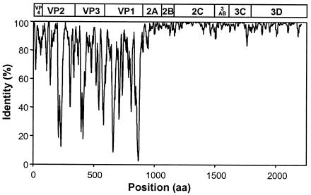
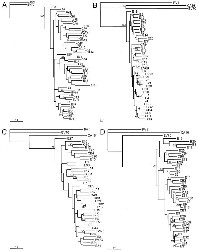
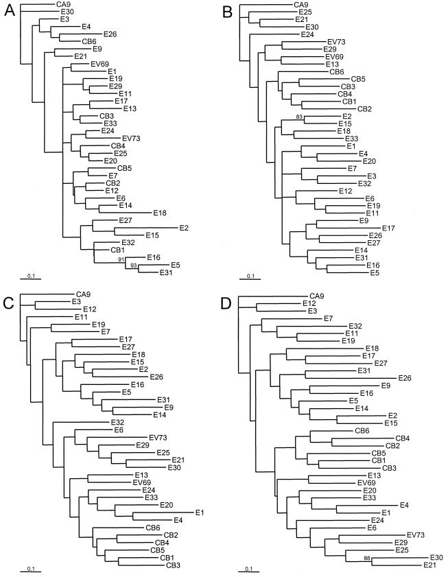
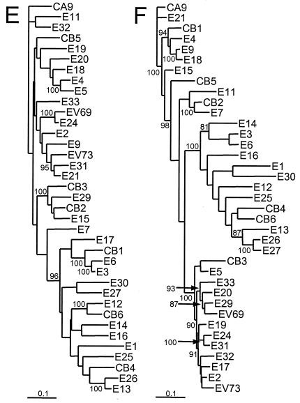
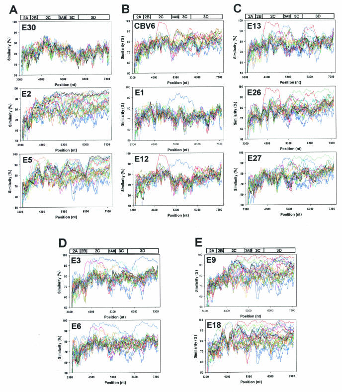
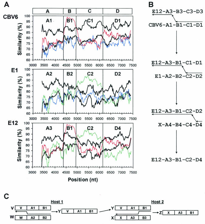
The species Human enterovirus B (HEV-B) in the family Picornaviridae consists of coxsackievirus A9; coxsackieviruses B1 to B6; echoviruses 1 to 7, 9, 11 to 21, 24 to 27, and 29 to 33; and enteroviruses 69 and 73. We have determined complete genome sequences for the remaining 22 HEV-B serotypes whose sequences were not represented in public databases and analyzed these in conjunction with previously available complete sequences in GenBank. Members of HEV-B were monophyletic relative to all other human enterovirus species in all regions of the genome except in the 5'-nontranslated region (NTR), where they are known to cluster with members of HEV-A. Within HEV-B, phylogenies constructed from the structural (P1) and nonstructural regions of the genome (P2 and P3) are incongruent, suggesting that recombination had occurred. Similarity plots and bootscanning analysis across the complete genome identified multiple sites at which the phylogeny of a given strain's sequence shifted, indicating potential recombination points. These points are distributed in the 5'-NTR and throughout P2 and P3, but no sites with >80% bootstrap support were identified within the capsid. Individual sequence comparisons and phylogenetic analyses suggest that members of HEV-B have recombined with one another on multiple occasions, resulting in a complex mosaic of sequences derived from multiple parental viruses in the nonstructural regions of the genome. We conclude that RNA recombination is a common mechanism for enterovirus evolution and that recombination within the nonstructural regions of the genome (P2 and P3) has been observed only among members of the same species.


Figures

FIG. 1.
FIG. 1.
Similarity plot summarizing sequence identities among HEV-B polyproteins. Identities among the 37 aligned sequences were plotted in the center of a 10-residue window, and the window was progressively advanced in one-residue steps. The enterovirus genetic map is shown at the top.
FIG. 2.
FIG. 2.
Phylogenetic trees based on HEV-B virus nucleotide sequences. Each of the major functional regions of the genome was analyzed independently. Bootstrap values (percentages of 1,000 pseudoreplicate data sets) supporting each cluster are shown at the nodes; for clarity, only values of > 80% are shown. CAV16, PV1, and EV70, representatives of HEV-A, HEV-C, and HEV-D, respectively, are included as outgroup taxa. All trees are plotted to the same scale, except for panel B (see scale bars). (A) 5′-NTR; (B) complete P1 region; (C) complete P2 region; (D) complete P3 region.
FIG. 3.
FIG. 3.
Phylogenetic trees based on HEV-B virus nucleotide sequences. The regions encoding each of the mature capsid proteins, the 2C protein, and the 3D protein were analyzed independently. Bootstrap values (percentages of 1,000 pseudoreplicate data sets) supporting each cluster are shown at the nodes; for clarity, only values of >80% are shown. All trees are plotted to the same scale (see scale bars). (A) 1A (VP4); (B) 1B (VP2); (C) 1C (VP3); (D) 1D (VP1); (E) 2C; (F) 3D.
FIG. 3.
FIG. 3.
Phylogenetic trees based on HEV-B virus nucleotide sequences. The regions encoding each of the mature capsid proteins, the 2C protein, and the 3D protein were analyzed independently. Bootstrap values (percentages of 1,000 pseudoreplicate data sets) supporting each cluster are shown at the nodes; for clarity, only values of >80% are shown. All trees are plotted to the same scale (see scale bars). (A) 1A (VP4); (B) 1B (VP2); (C) 1C (VP3); (D) 1D (VP1); (E) 2C; (F) 3D.
FIG. 4.
FIG. 4.
Representative similarity plots of HEV-B virus P2 and P3 nucleotide sequences calculated by SimPlot 3.2 beta (39). Each point represents the similarity between the query sequence and a given heterologous sequence, within a sliding window of 200 nucleotides centered on the position plotted, with a step of 20 residues between points. Positions containing gaps were excluded from the analysis. The enterovirus genetic map is shown at the top of each panel. For each plot, the identity of the query sequence is indicated in the upper left corner. Within panels B to E, homologous peaks are depicted in the same color. (A) E30-Bastianni, E2-Cornelis, and E5-Noyce; (B) CBV6-Schmitt, E1-Farouk, and E12-Travis; (C) E13-Del Carmen, E26-Coronel, and E27-Bacon; (D) E3-Morissey and E6-D'Amori; and (E) E9-Hill and E18-Metcalf.
FIG. 5.
FIG. 5.
Putative recombination pathways for CBV6-Schmitt, E1-Farouk, and E12-Travis, deduced from similarity plots and bootscanning analysis and a simple, schematic representation of recombination involving three hypothetical parental strains. (A) Similarity plots, as in Fig. 4, comparing the relationships among CBV6-Schmitt, E1-Farouk, and E12-Travis with their relationships to the other prototype strains. The curves depicting comparisons between CBV6, E1, and E12 are color-coded as follows: blue, CBV6-E1; red, CBV6-E12; and green, E1-E12. For each query sequence, the average similarity to strains other than CBV6-Schmitt, E1-Farouk, and E12-Travis is plotted in the lower black curve; the upper black curve indicates three standard deviations above the mean. The boundaries of regions A, B, C, and D, at the top, indicate sites where the relationships change. Within each of these regions, distinct alleles are labeled A1, A2, A3, etc. (B) Minimum recombination pathway to produce the observed virus isolates. Capsid identities are indicated by the serotype designations, CBV6, E1, E12, and X, where X is an unobserved strain of any HEV-B serotype, as described in the text. Regions and alleles are indicated as in panel A. Arrows pointing from right to left indicate minus strand synthesis and template switching to produce chimeric RNAs. (C) Coinfection of host 1 with strains V and W (recombination results in strain Y) and coinfection of host 2 with strains X and Y (recombination results in strain Z). Arrows between the parental strains indicate hypothetical template-switch points during minus strand synthesis.

References

    1. Andersson, P., K. Edman, and A. M. Lindberg. 2002. Molecular analysis of the echovirus 18 prototype: evidence of interserotypic recombination with echovirus 9. Virus Res. 85:71-83. - PubMed
    1. Blomqvist, S., A.-L. Bruu, M. Stenvik, and T. Hovi. 2003. Characterization of a recombinant type 3/type 2 poliovirus isolated from a healthy vaccinee and containing a chimeric capsid protein VP1. J. Gen. Virol. 84:573-580. - PubMed
    1. Branche, W. C., V. M. Young, F. M. Houston, and L. W. Koontz. 1965. Characterization of prototype virus ECHO-32. Proc. Soc. Exp. Biol. Med. 118:186-190. - PubMed
    1. Brown, B. A., K. Maher, M. S. Oberste, and M. A. Pallansch. 2003. Complete genomic sequencing shows that polioviruses and members of human enterovirus species C are closely related in the non-capsid coding region. J. Virol. 77:8973-8984. - PMC - PubMed
    1. Cammack, N., A. Phillips, G. Dunn, V. Patel, and P. D. Minor. 1988. Intertypic genomic rearrangements of poliovirus strains in vaccinees. Virology 167:507-514. - PubMed

LinkOut - more resources