

Differentiation stage-specific activation of p38 mitogen-activated protein kinase isoforms in primary human erythroid cells
- PMID: 14694199
- PMCID: PMC314153
- DOI: 10.1073/pnas.0307075101
Differentiation stage-specific activation of p38 mitogen-activated protein kinase isoforms in primary human erythroid cells
Abstract
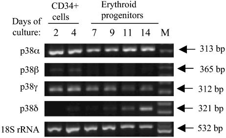
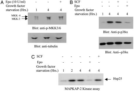
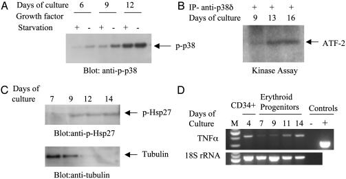
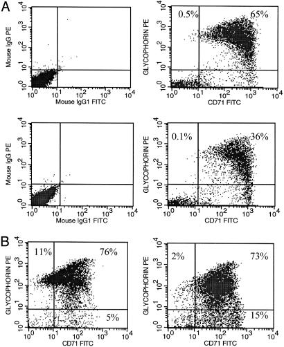
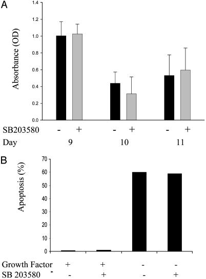
p38alpha, p38beta, p38gamma, and p38delta are four isoforms of p38 mitogen-activated protein (MAP) kinase (MAPK) involved in multiple cellular functions such as cell proliferation, differentiation, apoptosis, and inflammation response. In the present study, we examined the mRNA expression pattern of each of the four isoforms during erythroid differentiation of primary erythroid progenitors. We show that p38alpha and p38gamma transcripts are expressed in early hematopoietic progenitors as well as in late differentiating erythroblasts, whereas p38delta mRNA is only expressed and active during the terminal phase of erythroid differentiation. On the other hand, p38beta is minimally expressed in early CD34(+) hematopoietic progenitors but not expressed in lineage-committed erythroid progenitors. We also determined the phosphorylation/activation of p38alpha, MAPK kinase 3/6, and MAPKAP-2 in response to erythropoietin and stem cell factor. We found that phosphorylation of p38alpha, MAPK kinase kinase 3/6 and MAPKAP-2 occurs only upon growth factor withdrawal in primary erythroid progenitors. Moreover, our data indicate that activation of p38alpha does not induce apoptosis or promote proliferation of erythroid progenitors. On the other hand, under steady-state culture conditions, both p38alpha and p38delta isoforms are increasingly phosphorylated activated in the terminal phase of differentiation. This increased phosphorylation/activity was accompanied by up-regulation of heat shock protein 27 phosphorylation. Finally, we demonstrate that tumor necrosis factor alpha, an inflammatory cytokine that is modulated by p38alpha, is expressed by differentiating erythroblasts and inhibition of p38alpha or tumor necrosis factor alpha results in reduction in differentiation. Taken together, our data demonstrate that both p38alpha and delta isoforms function to promote the late-stage differentiation of primary erythroid progenitors and are likely to be involved in functions related to erythrocyte membrane remodeling and enucleation.
Figures
Similar articles
-
Selective activation and functional significance of p38alpha mitogen-activated protein kinase in lipopolysaccharide-stimulated neutrophils.J Clin Invest. 1999 Mar;103(6):851-8. doi: 10.1172/JCI5257. J Clin Invest. 1999. PMID: 10079106 Free PMC article.
-
Differential expression and activation of p38 mitogen-activated protein kinase alpha, beta, gamma, and delta in inflammatory cell lineages.J Immunol. 1999 Apr 1;162(7):4246-52. J Immunol. 1999. PMID: 10201954
-
Activation of discoidin domain receptor 1 facilitates the maturation of human monocyte-derived dendritic cells through the TNF receptor associated factor 6/TGF-beta-activated protein kinase 1 binding protein 1 beta/p38 alpha mitogen-activated protein kinase signaling cascade.J Immunol. 2003 Oct 1;171(7):3520-32. doi: 10.4049/jimmunol.171.7.3520. J Immunol. 2003. Retraction in: J Immunol. 2010 Aug 1;185(3):1984. doi: 10.4049/jimmunol.1090057. PMID: 14500648 Retracted.
-
Nuclear export of the stress-activated protein kinase p38 mediated by its substrate MAPKAP kinase-2.Curr Biol. 1998 Sep 24;8(19):1049-57. doi: 10.1016/s0960-9822(98)70442-7. Curr Biol. 1998. PMID: 9768359 Review.
-
p38 MAP-kinases pathway regulation, function and role in human diseases.Biochim Biophys Acta. 2007 Aug;1773(8):1358-75. doi: 10.1016/j.bbamcr.2007.03.010. Epub 2007 Mar 24. Biochim Biophys Acta. 2007. PMID: 17481747 Review.
Cited by
-
High resolution methylome analysis reveals widespread functional hypomethylation during adult human erythropoiesis.J Biol Chem. 2013 Mar 29;288(13):8805-14. doi: 10.1074/jbc.M112.423756. Epub 2013 Jan 10. J Biol Chem. 2013. PMID: 23306203 Free PMC article.
-
Methylation of dual-specificity phosphatase 4 controls cell differentiation.Cell Rep. 2021 Jul 27;36(4):109421. doi: 10.1016/j.celrep.2021.109421. Cell Rep. 2021. PMID: 34320342 Free PMC article.
-
Greensporone A, a Fungal Secondary Metabolite Suppressed Constitutively Activated AKT via ROS Generation and Induced Apoptosis in Leukemic Cell Lines.Biomolecules. 2019 Mar 29;9(4):126. doi: 10.3390/biom9040126. Biomolecules. 2019. PMID: 30934922 Free PMC article.
-
Alternative pre-mRNA splicing switches modulate gene expression in late erythropoiesis.Blood. 2009 Apr 2;113(14):3363-70. doi: 10.1182/blood-2008-05-160325. Epub 2009 Feb 4. Blood. 2009. PMID: 19196664 Free PMC article.
-
Erythrocyte mitogen-activated protein kinases mediate hemolytic events under osmotic and oxidative stress and in hemolytic diseases.Cell Signal. 2022 Nov;99:110450. doi: 10.1016/j.cellsig.2022.110450. Epub 2022 Aug 25. Cell Signal. 2022. PMID: 36029940 Free PMC article.
References
-
- Ono, K. & Han, J. (2000) Cell. Signaling 12, 1–13. - PubMed
-
- Shi, Y. & Gaestel, M. (2202) Biol. Chem. 383, 1519–1536. - PubMed
-
- Kessler, G. A., Bray, J., Hunt, J., Johnson, D. A., Gleason, T., Yao, Z., Wang, S.-W., Parker, C., Yamone, H., Cole, C. & Lichenstein, H. S. (1998) Express Purif. 14, 221–230. - PubMed
-
- Enslen, H., Raingeaud, J. & Davis, R. J. (1998) J. Biol. Chem. 273, 1741–1748. - PubMed
-
- Kumar, S., McDonnel, P. C., Gum, R. J., Hand, A. J. & Young, P. R. (1997) Biochem. Biophys. Res. Commun. 235, 533–538. - PubMed
Publication types
MeSH terms
Substances
LinkOut - more resources
Full Text Sources
Other Literature Sources
Research Materials
Miscellaneous
