. 2004 Feb 2;469(2):153-69.
doi: 10.1002/cne.11003.

Actions of a histaminergic/peptidergic projection neuron on rhythmic motor patterns in the stomatogastric nervous system of the crab Cancer borealis

Affiliations

Actions of a histaminergic/peptidergic projection neuron on rhythmic motor patterns in the stomatogastric nervous system of the crab Cancer borealis

Andrew E Christie et al. J Comp Neurol. .

Abstract

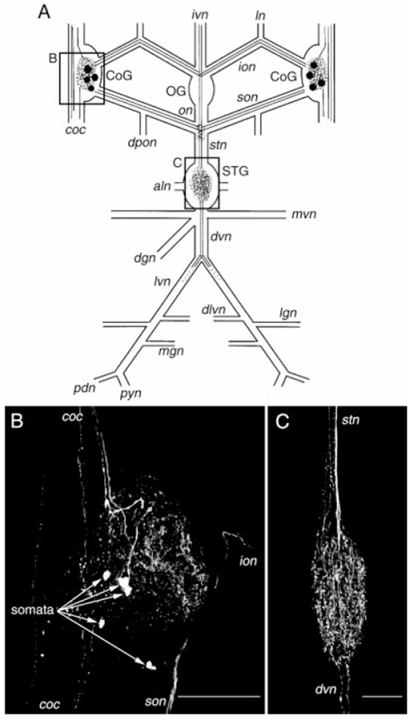
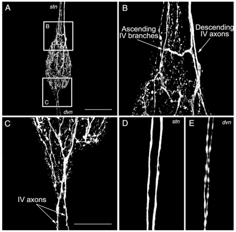
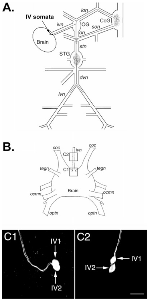
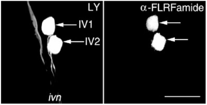
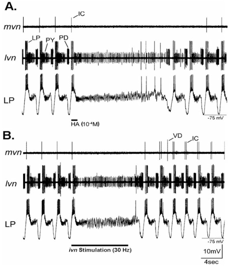
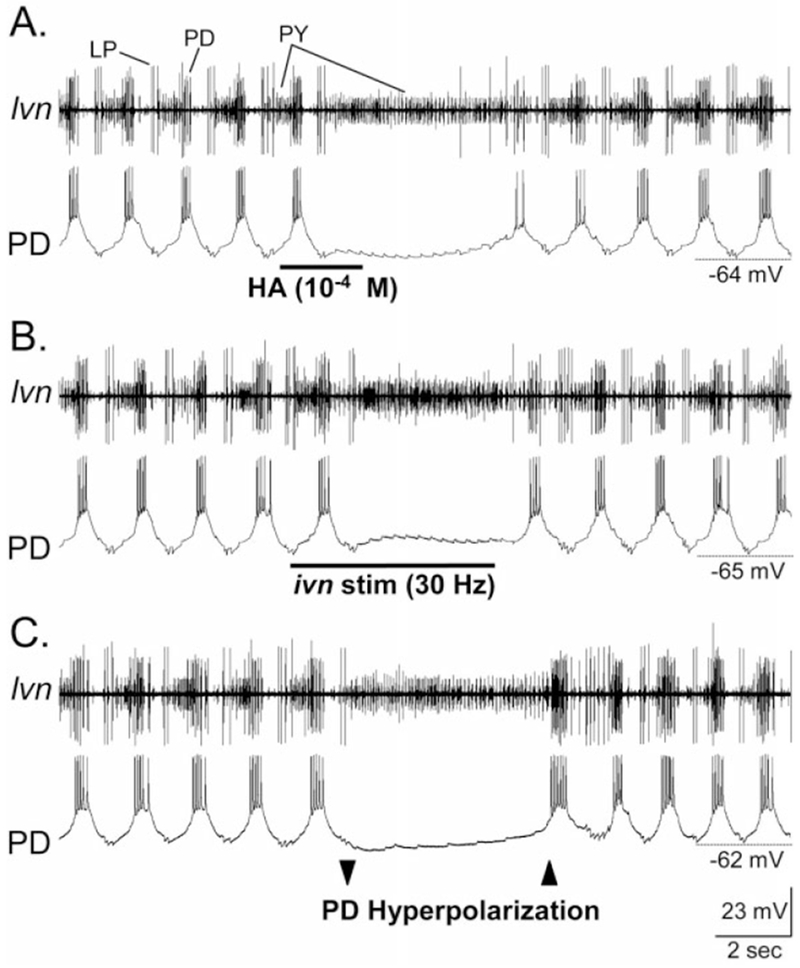
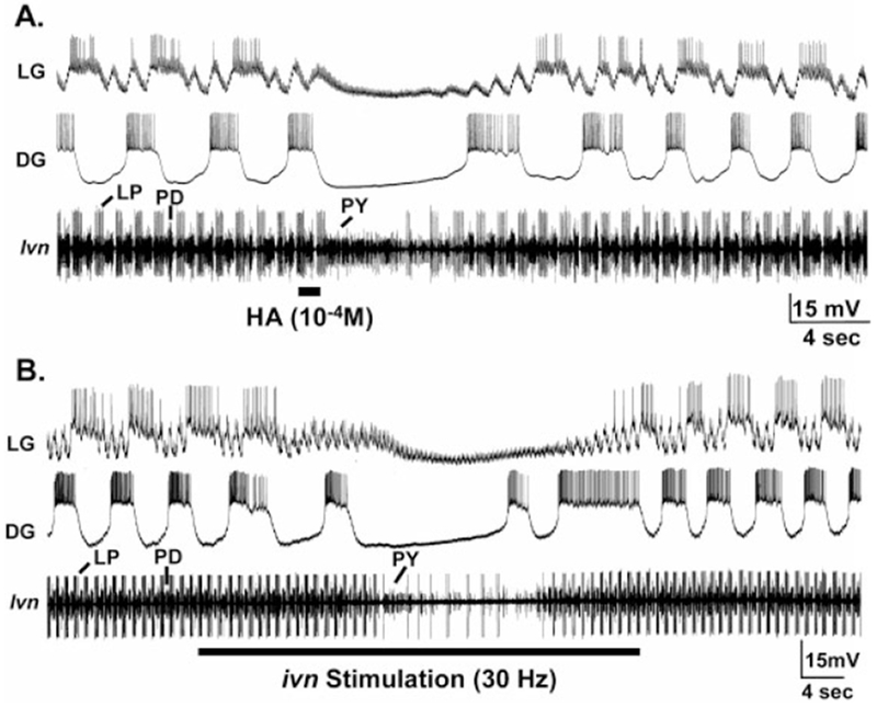
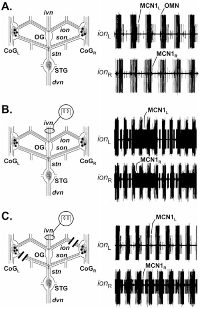
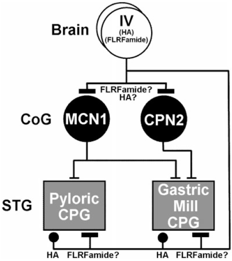
Histamine is a neurotransmitter with actions throughout the nervous system of vertebrates and invertebrates. Nevertheless, the actions of only a few identified histamine-containing neurons have been characterized. Here, we present the actions of a histaminergic projection neuron on the rhythmically active pyloric and gastric mill circuits within the stomatogastric ganglion (STG) of the crab Cancer borealis. An antiserum generated against histamine labeled profiles throughout the C. borealis stomatogastric nervous system. Labeling occurred in several somata and neuropil within the paired commissural ganglia as well as in neuropil within the STG and at the junction of the superior oesophageal and stomatogastric nerves. The source of all histamine-like immunolabeling in the STG neuropil was one pair of neuronal somata, the previously identified inferior ventricular (IV) neurons, located in the supraoesophageal ganglion. These neurons also exhibited FLRFamide-like immunoreactivity. Activation of the IV neurons in the crab inhibited some pyloric and gastric mill neurons and, with inputs from the commissural ganglia eliminated, terminated both rhythms. Focal application of histamine had comparable effects. The actions of both applied histamine and IV neuron stimulation were blocked, reversibly, by the histamine type-2 receptor antagonist cimetidine. With the commissural ganglia connected to the STG, IV neuron stimulation elicited a longer-latency activation of commissural projection neurons which in turn modified the pyloric rhythm and activated the gastric mill rhythm. These results support the hypothesis that the histaminergic/peptidergic IV neurons are projection neurons with direct and indirect actions on the STG circuits of the crab C. borealis.


Figures

Fig. 1.
Fig. 1.
Distribution of histamine-like (HA-like) immunolabeling in the stomatogastric nervous system (STNS) of Cancer borealis. A: Schematic representation of the STNS showing the distribution of HA-like labeling. Labeled somata are indicated by filled circles, axons by solid or dotted lines, and neuropil by stippling. The boxed areas of the schematic show the relative positions of the photomicrographs shown in B and C. B: Confocal photomicrograph showing an HA-like immunolabeled commissural ganglion (CoG). In this image, five somata are present (arrows) as well as neuropil and axons in several of the nerves that connect with this ganglion. C: Confocal photomicrograph showing the distribution of HA-like immunolabeling in the stomatogastric ganglion (STG). HA-like staining within the STG is restricted to the neuropil. None of the approximately 25 neurons present in this ganglion exhibit HA-like immunoreactivity. B and C are composite images of 41 and 28 optical sections, respectively, taken at approximately 2μm intervals. Ganglia: OG, oesophageal ganglion. Nerves: aln, anterior lateral nerve; coc, circumoesophageal connective; dgn, dorsal gastric nerve; dlvn, dorsal lateral ventricular nerve; dpon, dorsal posterior oesophageal nerve; dvn dorsal ventricular nerve; ion, inferior oesophageal nerve; ivn, inferior ventricular nerve; lgn, lateral gastric nerve; ln, labral nerve; lvn, lateral ventricular nerve; mgn, medial gastric nerve; mvn, medial ventricular nerve; on, oesophageal nerve; pdn, pyloric dilator nerve; pyn, pyloric nerve; stn, stomatogastric nerve; son, superior oesophageal nerve. Scale bars = 250 μm in B, 100 μm in C.
Fig. 2.
Fig. 2.
Branching pattern of the histaminergic projections in the stomatogastric ganglion. A: Composite image of HA-like immunolabeling in the STG. Note the presence of multiple fibers where the stomatogastric nerve (stn) and the dorsal ventricular nerve (dvn) connect with the STG. The boxed areas represented magnified regions shown in B and C. B: The HA-like immunolabeling of fibers at the junction of the stn and STG represents both relatively small diameter ascending fibers that extend no further than halfway along the stn and two larger diameter descending fibers that project from the IV neurons in the brain to the STG neuropil. C: In some STGs, HA-like immunolabeling projects past the STG in the dvn. D,E: The descending stn (D) and dvn (E) HA-like immunolabeled fibers exhibit different morphology. The descending stn fibers have a relatively large diameter and appear to be fibers of passage. The dvn fibers have a smaller diameter and a beads on a string morphology, comparable to that of the fibers ascending from the STG. The descending stn fibers could be traced directly from the immunolabeled fibers in the ivn. The image in D was collected in the anterior region of the stn, at a point anterior to the region containing the smaller immunolabeled fibers that ascend from the STG. The images in A-E are composites of 34, 26, 23, 19, and 12 optical sections, respectively. The optical sections were collected at 2-μm intervals in A and 1-μm intervals in B–E. Scale bars = 250 μm in A, 100 μm in C (applies to B–E).
Fig. 3.
Fig. 3.
Soma location and projection pattern of the inferior ventricular (IV) neurons in Cancer borealis. A: Schematic representation of the stomatogastric nervous system connected with the supraoesophageal ganglion (Brain), showing the projection pattern of one of the paired IV neurons. These two neurons have identical projection patterns. The IV neuron somata are indicated by filled circles, and the axonal projection pattern is represented by solid lines. The neuropilar areas of the IV neurons are indicated by stippling. B: Schematic representation of the brain of C. borealis with some of its major nerves. The boxed regions in this schematic show the location of the confocal photomicrographs of HA-immunolabeled preparations shown in C1 and C2. C: C1 shows the IV neurons (IV1, IV2) in their more common location, within the brain at the insertion point of the inferior ventricular nerve (ivn). C2 shows the IV neurons within the ivn, where it occurred in fewer than 10% of the preparations examined. C1 and C2, which were taken from different preparations, are composite images of 25 and 16 optical sections, respectively. Images for both panels were collected at approximately 2-μm intervals and are shown at the same scale. ocmn, oculomotor nerve; optn, optic nerve; tegn, tegumentary nerve. For other abbreviations, see Figure 1. Scale bar = 50 μm in C2 (applies to C1,C2).
Fig. 4.
Fig. 4.
The IV neurons of Cancer borealis contain a FLRFamide-like peptide cotransmitter. Lucifer yellow (LY) nerve backfills of the inferior ventricular nerve (ivn) were paired with anti-FLRFamide immunoprocessing to show that the IV neurons exhibit FLRFamide-like immunoreactivity. Left: LY backfill of the ivn resulted in LY labeling in the brain, near the insertion point of the ivn, of only the two IV neuron somata (arrows). Right: The distribution of FLRFamide-like immunolabeling, in the same region of the brain from the same preparation as the LY backfill panel, visualized by means of a rhodamine-conjugated secondary antibody. The two IV neurons (arrows) exhibit FLRFamide-like immunolabeling. Each panel is a composite of 31 optical sections taken at approximately 1-μm intervals that were collected simultaneously. Scale bar = 100 μm.
Fig. 5.
Fig. 5.
Inhibition of the pyloric rhythm by HA application and IV neuron stimulation in preparations where the STG was isolated from the commissural ganglia. A: Focal application of HA (bar) onto the STG neuropil caused a long-lasting but reversible suppression of the pyloric rhythm. Note the loss of rhythmic neuronal activity in both extracellular recordings (mvn, lvn) and in the intracellular recording of the lateral pyloric (LP) neuron. During this time, only PY neuron activity persisted (lvn), although the LP neuron fired a few action potentials just before the pyloric rhythm resumed. The inhibitory postsynaptic potentials (IPSPs) that occurred in the LP neuron while the pyloric rhythm was turned off represent input from the PY neurons (Selverston and Moulins, 1987). In this preparation, there was no ongoing VD neuron activity after the STG was isolated. B: Stimulation of the ivn (30 Hz) reversibly terminated the ongoing pyloric rhythm. This rhythm resumed approximately 2 seconds after the stimulation was stopped. Comparable to the pyloric rhythm response to HA application, the persisting tonic activity in the lvn recording during ivn stimulation was from the PY neurons. As in A, the PY neurons were responsible for the persisting IPSPs that occurred in the LP neuron during nerve stimulation. A and B are from the same preparation. For abbreviations, see list. Scale bars are the same for both panels.
Fig. 6.
Fig. 6.
The pyloric circuit response to HA application and ivn stimulation is similar to that resulting from direct hyperpolarization of the pyloric pacemaker neurons. A: Focal application of HA onto the STG neuropil caused a long-lasting but reversible suppression of the pyloric rhythm (lvn). Tonic PY neuron activity (lvn) was all that persisted during pyloric rhythm suppression. Note that the PD neuron membrane potential remained hyperpolarized in response to the HA application. There are two PD neurons, both of which are members of the electrically coupled, pyloric pacemaker ensemble. B: IV neuron stimulation terminated the ongoing pyloric rhythm. As occurred in response to HA application, only PY neuron activity persisted and the PD neuron remained at a hyperpolarized membrane potential. C: Intracellular hyperpolarizing current injection into a PD neuron terminated the ongoing pyloric rhythm (lvn). As occurred in response to HA application and ivn stimulation, only the PY neurons remained active during the PD neuron hyperpolarization. In this experiment, one PD neuron was injected with hyperpolarizing current (not shown), while the second PD neuron was simultaneously recorded. All three panels are from the same preparation, in which the STG was isolated from the CoGs. For abbreviations, see list. Scale bars are the same for all panels.
Fig. 7.
Fig. 7.
Inhibition of the gastric mill rhythm by histamine (HA) application and IV neuron stimulation in preparations where the STG was isolated from the commissural ganglia. A: Focal application of HA onto the STG neuropil reversibly terminated the gastric mill (LG, DG) and pyloric rhythms (lvn). The gastric mill rhythm was driven, and the pyloric rhythm modulated, by continuous stimulation (15 Hz each) of both MCN1 projection neurons for the duration of the recording. The persisting excitatory postsynaptic potentials (EPSPs) in the LG neuron during ivn stimulation represent electrical EPSPs from MCN1 (Coleman et al., 1995). Most hyperpolarized Vm: DG, −68 mV; LG, −76 mV. B: Extracellular stimulation of the ivn (30 Hz) reversibly terminated ongoing gastric mill (LG, DG) and pyloric rhythms (lvn). As in A, both MCN1s were stimulated tonically (15 Hz each) for the duration of the recording, and the electrical EPSPs from MCN1 in the LG neuron persisted during the gastric mill response to HA application. Most hyperpolarized Vm: DG, −68mV; LG, −74 mV. A and B are from the same preparation. For abbreviations, see list.
Fig. 8.
Fig. 8.
IV neuron stimulation activates the gastric mill rhythm, strengthens the pyloric and oesophageal rhythms, and excites CoG projection neurons when the STG remains connected with the CoGs. Left: Before ivn stimulation, there was no gastric mill rhythm (GM, LG, dgn, mvn), relatively weak pyloric (mvn) and oesophageal (ion) rhythms, and little spontaneous activity in the MCN1 projection neuron (ion). The oesophageal rhythm is monitored by rhythmic bursting of an oesophageal motor neuron (OMN: ion). The tonically active unit in the dgn represents the activity of the anterior gastric receptor (AGR) sensory neuron, which is commonly tonically active in the isolated stomatogastric nervous system. Right: After ivn stimulation (30 Hz), the gastric mill rhythm was elicited and the pyloric and oesophageal rhythms were strengthened. Note that there was gastric mill-timed inhibition of the pyloric-timed activity in the IC and VD neurons (mvn), as commonly occurs during some versions of the gastric mill rhythm (Blitz and Nusbaum, 1997). Similarly, MCN1 (ion) exhibited both a gastric mill-timed (during the LG/GM neuron burst) and pyloric-timed (during the DG neuron burst) activity pattern. For other abbreviations, see list.
Fig. 9.
Fig. 9.
Only one of the two IV neuron projections to each CoG excites the MCN1 projection neuron. A: Simultaneous extracellular recordings of the left (ionL) and right (ionR) ions before ivn stimulation showed an ongoing oesophageal rhythm (OMN) and weak oesophageal-timed activity from both MCN1s. B: After ivn stimulation, both MCN1s exhibited strengthened activity that was now time-locked with both the pyloric rhythm (short duration bursts) and the gastric mill rhythm (long duration bursts) (see Fig. 8). C: After transection of the sonL and ionR, ivn stimulation no longer excited MCN1L but it still excited MCN1R. This result indicated that IV neuron excitation of MCN1 occurs only by means of the son axon of the IV neuron. Note that the ionR was transected on the STG side of the ionR recording electrode. This method enabled continued recording of MCN1 activity projecting from the CoG, but it prevented this MCN1 activity from reaching the STG. No gastric mill rhythm was activated, because MCN1L was not excited and MCN1R activity did not reach the STG. The recordings in all three panels are from the same preparation.
Fig. 10.
Fig. 10.
Schematic of the direct and indirect actions of the IV neurons on the gastric mill and pyloric central pattern generating circuits (CPGs) in the STG of the crab Cancer borealis. The IV neurons use the neurotransmitter histamine (HA) to elicit a direct, relatively short-lasting inhibition of both of these CPGs. It remains to be determined whether there is also a peptidergic (FLRFamide-like) component to the IV neuron actions in the STG. By means of their excitation of CoG projection neurons, including MCN1 and CPN2, the IV neurons also elicit an indirect, relatively long-lasting excitation of the gastric mill and pyloric CPGs. The IV neuron excitation of these projection neurons may result from the actions of its peptide cotransmitter. For other abbreviations, see list. Symbols: T-bars, excitation; Filled circles, inhibition.

Similar articles

Cited by

References

    1. Bartos M, Nusbaum MP. 1997. Intercircuit control of motor pattern modulation by presynaptic inhibition. J Neurosci 17:2247–2256. - PMC - PubMed
    1. Bartos M, Manor Y, Nadim F, Marder E, Nusbaum MP. 1999. Coordination of fast and slow rhythmic neuronal circuits. J Neurosci 19:6650–6660. - PMC - PubMed
    1. Battelle BA, Calman BG, Andrews AW, Grieco FD, Mleziva MB, Callaway JC, Stuart AE. 1991. Histamine: a putative afferent neurotransmitter in Limulus eyes. J Comp Neurol 305:527–542. - PubMed
    1. Battelle BA, Calman BG, Hart MK. 1999. Cellular distributions and functions of histamine, octopamine, and serotonin in the peripheral visual system, brain, and circumoesophageal ring of the horseshoe crab Limulus polyphemus. Microsc Res Tech 44:70–80. - PubMed
    1. Blitz DM, Nusbaum MP. 1997. Motor pattern selection via inhibition of parallel pathways. J Neurosci 17:4965–4975. - PMC - PubMed

Publication types

MeSH terms

LinkOut - more resources