. 2004 Jan;86(1 Pt 1):428-34.
doi: 10.1016/S0006-3495(04)74119-3.

Increasing the amphiphilicity of an amyloidogenic peptide changes the beta-sheet structure in the fibrils from antiparallel to parallel

Affiliations

Increasing the amphiphilicity of an amyloidogenic peptide changes the beta-sheet structure in the fibrils from antiparallel to parallel

David J Gordon et al. Biophys J. 2004 Jan.

Abstract

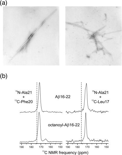
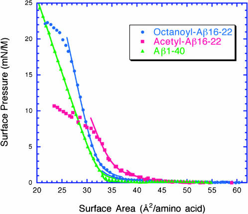
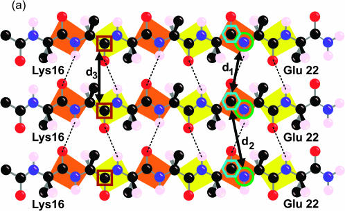
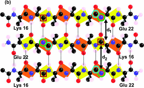
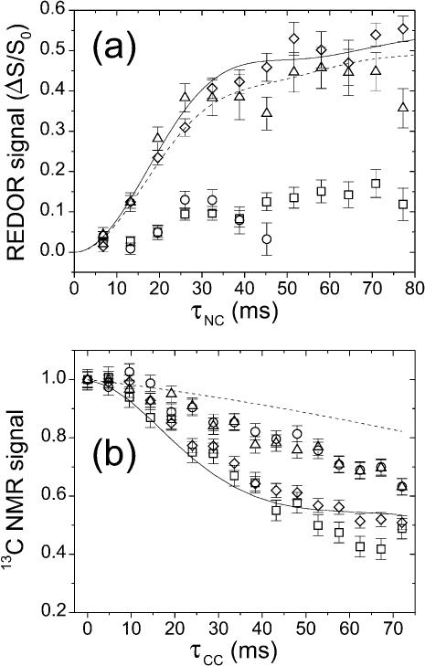
Solid-state NMR measurements have been reported for four peptides derived from beta-amyloid peptide Abeta(1-42): Abeta(1-40), Abeta(10-35), Abeta(16-22), and Abeta(34-42). Of these, the first two are predicted to be amphiphilic and were reported to form parallel beta-sheets, whereas the latter two peptides appear nonamphiphilic and adopt an antiparallel beta-sheet organization. These results suggest that amphiphilicity may be significant in determining fibril structure. Here, we demonstrate that acylation of Abeta(16-22) with octanoic acid increases its amphiphilicity and changes the organization of fibrillar beta-sheet from antiparallel to parallel. Electron microscopy, Congo Red binding, and one-dimensional 13C NMR measurements demonstrate that octanoyl-Abeta(16-22) forms typical amyloid fibrils. Based on the stability of monolayers at the air-water interface, octanoyl-Abeta(16-22) is more amphiphilic than Abeta(16-22). Measurements of 13C-13C and 15N-13C nuclear magnetic dipole-dipole couplings in isotopically labeled fibril samples, using the constant-time finite-pulse radiofrequency-driven recoupling (fpRFDR-CT) and rotational echo double resonance (REDOR) solid-state NMR techniques, demonstrate that octanoyl-Abeta(16-22) fibrils are composed of parallel beta-sheets, whereas Abeta(16-22) fibrils are composed of antiparallel beta-sheets. These data demonstrate that amphiphilicity is critical in determining the structural organization of beta-sheets in the amyloid fibril. This work also shows that all amyloid fibrils do not share a common supramolecular structure, and suggests a method for controlling the structure of amyloid fibrils.


Figures

FIGURE 1
FIGURE 1
(a) Electron micrographs of negatively stained fibrils formed by the Aβ(16–22) and octanoyl-Aβ(16–22) peptides over a 14-day incubation. Magnification, 45,000×. Fibrils formed by both peptides exhibit the long, unbranched, and twisting morphology that is typical of amyloid fibrils. This demonstrates that the addition of the octanoyl group to Aβ(16–22) does not significantly alter the structure of the amyloid fibril on the macromolecular level. (b) Carbonyl regions of 1D 13C MAS NMR spectra of Aβ(16–22) and octanoyl-Aβ(16–22) fibrils with the indicated isotopic labels. Dashed lines indicate random coil chemical shift values for the carbonyl 13C labels. Experimental chemical shifts and linewidths in all spectra are consistent with well-structured β-strand conformations.
FIGURE 2
FIGURE 2
Surface isotherms of Aβ(16–22) (•), octanoyl-Aβ(16–22) (▪), and Aβ(1–40) (▴) peptide monolayers spread on a subphase of 25 mM phosphate buffer, 150 mM NaCl, 5% glycerol, pH 7.4. Data were fit to the equations described in Methods. Octanoyl-Aβ(16–22) and Aβ(1–40) form more stable monolayers than Aβ(16–22) based on the collapse pressures of the peptide monolayers. This demonstrates that the octanoyl group increases the amphiphilicity of octanoyl-Aβ(16–22) relative to Aβ(16–22).
FIGURE 3
FIGURE 3
Labeling scheme for REDOR and fpRFDR-CT solid-state NMR measurements designed to distinguish in-register parallel (a) from in-register antiparallel (b) β-sheet structures. Carbonyl 13C labels at Leu17 and Phe20 and amide 15N labels at Ala21 are indicated by brown square, cyan hexagon, and green circle outlines, respectively. The figure shows only the carbonyl and the β-carbon atom of the N-terminal acyl group, which is an acetyl group in Aβ(16–22) and an octanoyl group in octanoyl-Aβ(16–22). The figure shows the hydrogen atom on the α-carbon atom and only the β-carbon of the side chains.
FIGURE 3
FIGURE 3
Labeling scheme for REDOR and fpRFDR-CT solid-state NMR measurements designed to distinguish in-register parallel (a) from in-register antiparallel (b) β-sheet structures. Carbonyl 13C labels at Leu17 and Phe20 and amide 15N labels at Ala21 are indicated by brown square, cyan hexagon, and green circle outlines, respectively. The figure shows only the carbonyl and the β-carbon atom of the N-terminal acyl group, which is an acetyl group in Aβ(16–22) and an octanoyl group in octanoyl-Aβ(16–22). The figure shows the hydrogen atom on the α-carbon atom and only the β-carbon of the side chains.
FIGURE 4
FIGURE 4
(a) 13C-detected 15N-13C REDOR measurements on Aβ(16–22) and octanoyl-Aβ(16–22) fibrils. Data are shown for Aβ(16–22)-15N-Ala21:Aβ(16–22)-13C-Leu17 (▵), Aβ(16–22)-15N-Ala21:Aβ(16–22)-13C-Phe20 (○), octanoyl-Aβ(16–22)-15N-Ala21:octanoyl-Aβ(16–22)-13C-Leu17 (□), and octanoyl-Aβ(16–22)-15N-Ala21:octanoyl-Aβ(16–22)-13C-Phe20 (⋄) fibrils. Error bars represent uncertainty from the root mean-square (rms) noise in the NMR spectra. Simulated REDOR curves assume an in-register antiparallel β-sheet structure in Aβ(16–22) (dashed line) and an in-register parallel β-sheet structure in octanoyl-Aβ(16–22) (solid line). (b) 13C-13C fpRFDR-CT measurements on Aβ(16–22) and octanoyl-Aβ(16–22) fibrils (same symbols). Simulated curves assume an in-register antiparallel β-sheet structure in Aβ(16–22) (dashed line) and an in-register parallel β-sheet structure in octanoyl-Aβ(16–22) (solid line).

Comment in

References

    1. Aggeli, A., M. Bell, N. Boden, J. N. Keen, P. F. Knowles, T. C. McLeish, M. Pitkeathly, and S. E. Radford. 1997. Responsive gels formed by the spontaneous self-assembly of peptides into polymeric β-sheet tapes. Nature. 386:259–262. - PubMed
    1. Anderson, R. C., T. Gullion, J. M. Joers, M. Shapiro, E. B. Villhauer, and H. P. Weber. 1995. Conformation of [1-13C,15N]acetyl-L-carnitine. Rotational-echo double resonance nuclear magnetic resonance spectroscopy. J. Am. Chem. Soc. 117:10546–10550.
    1. Antzutkin, O. N., J. J. Balbach, R. D. Leapman, N. W. Rizzo, J. Reed, and R. Tycko. 2000. Multiple quantum solid-state NMR indicates a parallel, not antiparallel, organization of β-sheets in Alzheimer's β-amyloid fibrils. Proc. Natl. Acad. Sci. USA. 97:13045–13050. - PMC - PubMed
    1. Balbach, J. J., Y. Ishii, O. N. Antzutkin, R. D. Leapman, N. W. Rizzo, F. Dyda, J. Reed, and R. Tycko. 2000. Amyloid fibril formation by Aβ 16–22, a seven-residue fragment of the Alzheimer's β-amyloid peptide, and structural characterization by solid state NMR. Biochemistry. 39:13748–13759. - PubMed
    1. Balbach, J. J., A. T. Petkova, N. A. Oyler, O. N. Antzutkin, D. J. Gordon, S. C. Meredith, and R. Tycko. 2002. Supramolecular structure in full-length Alzheimer's β-amyloid fibrils: evidence for a parallel β-sheet organization from solid state NMR. Biophys. J. 83:1205–1216. - PMC - PubMed

Publication types

LinkOut - more resources