Comparative Study
. 2004 Jan;164(1):91-100.
doi: 10.1016/S0002-9440(10)63100-8.

K-ras gene mutation enhances motility of immortalized airway cells and lung adenocarcinoma cells via Akt activation: possible contribution to non-invasive expansion of lung adenocarcinoma

Affiliations
Comparative Study

K-ras gene mutation enhances motility of immortalized airway cells and lung adenocarcinoma cells via Akt activation: possible contribution to non-invasive expansion of lung adenocarcinoma

Koji Okudela et al. Am J Pathol. 2004 Jan.

Abstract

Point mutations of the K-ras gene, which are found in 10 to 30% of lung adenocarcinomas, are regarded as being an early event during the carcinogenesis. Autonomous vigorous motility of neoplastic cells, as well as growth and survival advantages, are considered to be necessary for cancer development and progression. The present study describes the contributions of the K-ras gene mutation and its downstream pathway via phosphatidylinositol 3-OH kinase (PI3K)-Akt to the cell motility in an immortalized human peripheral airway epithelial cell (HPL1D) and lung adenocarcinoma cells (A549, H820, TKB6, and TKB14). We have also evaluated the relationship between pathological events and the K-ras-Akt pathway using surgically resected lung tumors. The HPL1D cells transfected with the mutated K-ras gene (HPL-V12) showed a significant increase in cell motility compared to those transfected with empty vector (HPL-E) or wild-type K-ras gene (HPL-K). The enhanced motility in the HPL-V12 cells was markedly reduced by either treatment with inhibitors of ras, PI3K, and/or MEK, or by transfection with the dominant-negative mutant Akt (dnAkt). The lung adenocarcinoma cells bearing the K-ras gene mutation (A549 and H820) showed consistently higher levels of cell motilities than those without the mutation (TKB6 and TKB14), and the motility of A549 and H820 cells were significantly inhibited by dnAkt transfection. These results suggest that the K-ras gene mutation could enhance the motility of neoplastic cells through a pathway involving PI3K-Akt. Actually, among the surgically resected lung tumors, the adenocarcinomas with the K-ras gene mutation tended to show a higher frequency and intensity of immunoreactivity for phosphorylated Akt (p-ser473Akt) than those without the mutation, supporting the in vitro observation that the mutated K-ras can activate the PI3K-Akt pathway. Immunoreactivity for p-ser473Akt was also seen in the pre-malignant and early lesions at a frequency similar to that in the advanced lung adenocarcinomas,. No correlation was seen between p-ser473Akt immunoreactivity and lymphatic/organ metastasis or prognosis. These results taken together suggest that the K-ras-Akt pathway might facilitate the motility of neoplastic cells during the early period of carcinogenesis in lung adenocarcinomas, and may contribute to their non-invasive expansion along the alveolar septa, rather than invasion or metastasis.


Figures
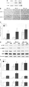
Figure 1
Figure 1
A: Expression of K-ras and c-met in mock (HPL-E), K-ras (HPL-K) and K-rasV12 (HPL-V12) transfectants. B: Autonomous migrations of the transfectants. The cells that migrated through the pores of the transwell membranes were stained with hematoxylin; magnification, ×100. C: The results of assays for autonomous and HGF-induced migration. Three independent assays were performed. The mean values of the ratio of stimulated to the autonomous migration of HPL-E cells (HGF (−)) are plotted. #/*; P <0.05. D: Phosphorylation status of Akt and Erk1/2. The transfectants were treated with (HGF) or without (Ctl) 20 ηg/ml HGF for 90 minutes. The protein extracts were resolved by SDS-PAGE, and blotted with antibodies against p-ser437Akt, p-Erk1/2, Akt, Erk1/2, or β-actin. E: Relative expression levels of p-ser473Akt (top) and p-Erk (Erk1, middle; Erk2, bottom). Three independent assays were performed. The expression levels were analyzed as described in Materials and Methods. The mean values of the expression level relative to that of HPL-E cells (HGF (−)) were plotted. #/*; P <0.05. Bar, SD.
Figure 2
Figure 2
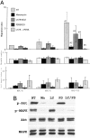
A: The effects of inhibitors of ras (manumycin, 50 μmol/L, Mn), PI3K (LY294002, 10 μmol/L, LY), and MEK (PD98059, 50 μmol/L, PD) on the autonomous migration (top), cell growth (middle), and cell death induction (bottom). Three independent assays were performed. The mean values of the migration or growth levels relative to those of HPL-E without treatment (NT), and cell death index (%) were plotted. #1/#2, #1/#5; P <0.05. Bar, SD. B: Effects of the inhibitors on phosphorylation status of Akt and Erk1/2. The protein extracts from the K-rasV12 cells treated or not with these inhibitors were resolved by SDS-PAGE, and blotted with antibodies against p-ser473Akt, p-Erk1/2, Akt, or Erk1/2.
Figure 3
Figure 3
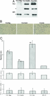
A: The protein extracts were resolved by SDS-PAGE, and blotted with antibodies against polyhistidine (His) or Akt. The faster migrating bands (#) are endogenous Akt, and the slower migrating bands (*) are exogenously expressed His-tagged Akt. V12/Emp, HPL-V12 transfected with the empty vector; V12/dnAkt, HPL-V12 with dominant-negative Akt; V12/Akt, HPL-V12 with wild-type Akt. B: Autonomous migration of V12/Emp, V12/dnAkt, and V12/Akt. The cells that migrated through the pores of the membranes were stained with hematoxylin; magnification ×100. C: The cell migration (top), cell growth (middle), and cell death (bottom) of the trasfectants. Three independent experiments were performed. The mean values of the ratio against the autonomous migration or growth value relative to those of HPL-E cells (described in Figure 1), and cell death index (%) are plotted. Bar, SD. #1/#2, #2/#3; P <0.05.
Figure 4
Figure 4
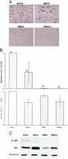
A: Autonomous migration of the lung adenocarcinoma cells with the K-ras gene mutation (A549 and H820) and those without the mutation (TKB6 and TKB14). The cells that migrated through the membranes were stained with hematoxylin; magnification ×100. B: The autonomous migration (top) and cell growth (bottom) of the cell lines. Three independent experiments were performed. The mean values of the autonomous migration or growth value relative to those of A549 cells are plotted. Bar, SD. #1/#3, #1/#4, #2/#3, #2/#4; P <0.05. C: Phosphorylation status of Akt in the cell lines. The protein extracts were immunoblotted with antibodies against p-ser473Akt, Akt, or β-actin.
Figure 5
Figure 5
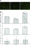
Effects of Akt/;dnAkt on autonomous migration, growth, and apoptosis in A549 cells. The desired genes inserted into pIRES-EGFP vector (10 μg) were transfected into A549 cells by using lipofectamine plus reagent. After 18 hours, the cells were trypsinized, and 5 × 10⁵ cells were subjected to the migration assay. The same numbers of cells were seed onto 6-well plates to examine the influence of Akt and dnAkt on cell growth. A part of the cell suspension was centrifuged using cytospin (Sakura, Tokyo, Japan) to mount cells on glass slides, and the cells were stained with 4,6-diamio-2-phenyliodole (DAPI). Under fluorescent microscopic observation, cells expressing EGFP were counted. Transfection efficacies were about 5 to 10%, and were not different among the mock, Akt, and dnAkt groups (data not shown). The cells that migrated through the pores of the membranes were observed by fluorescence microscopy, after 18 hours of incubation (A). The number of cells that expressed EGFP and passed through the membrane pores were counted and standardized by the transfection efficiency, and taken as migration values. Migration ratio (/mock cells) is plotted (B, top). The number of cells expressing EGFP in the 6-well plate after 18 hours of incubation were counted and standardized by the transfection efficiency, and were taken as growth values. Growth ratio (/mock cells) is plotted (B, middle). The percentage of cells showing the characteristics of apoptosis (nuclear condensation or fragmentation) among EGFP-expressing cells was determined as an apoptotic index (B, bottom). Bar, SD. #1/#2; P <0.05.
Figure 6
Figure 6
A: Immunohistochemistry for p-ser473Akt and Akt. A positive signal for Akt was observed in the cytoplasm of both cases with (case 55) and without (case 39) K-ras gene mutations. Diffuse strong immunoreactivity for p-ser473Akt could be seen in case 55, but not in case 39. B: P-ser473Akt expression and prognosis. Seventy-two cases of T1 (TNM classification) were selected and divided into two groups; cases with low (score <2.0, dashed line, 49 cases) and cases with high (score ≥2.0, solid line, 23 cases) immunoreactivity. Kaplan-Meier survival analysis was performed (P = 0.4307).

References

    1. Bos JL, Fearon ER, Hamilton SR, Verlaan-de Vries M, van Boom JH, van der Eb AJ, Vogelstein B. Prevalence of ras gene mutations in human colorectal cancers. Nature. 1987;327:293–297. - PubMed
    1. Bos JL, Verlaan-de Vries M, van der Eb AJ, Janssen JW, Delwel R, Lowenberg B, Colly LP. Mutations in N-ras predominate in acute myeloid leukemia. Blood. 1987;69:1237–1241. - PubMed
    1. Segrelle C, Ruiz S, Perez P, Murga C, Santos M, Budunova IV, Martinez J, Larcher F, Slaga TJ, Gutkind JS, Jorcan JL, Paramio JM. Functional roles of Akt signaling in mouse skin tumorigenesis. Oncogene. 2002;21:53–64. - PubMed
    1. Rodenhuis S, van de Wetering ML, Mooi WJ, Evers SG, van Zandwijk N, Bos JL. Mutational activation of the K-ras oncogene: a possible pathogenetic factor in adenocarcinoma of the lung. N Engl J Med. 1987;317:929–935. - PubMed
    1. Cooper CA, Carby FA, Bubb VJ, Lamb D, Kerr KM, Wyllie AH. The pattern of K-ras mutation in pulmonary adenocarcinoma defines a new pathway of tumor development in the human lung. J Pathol. 1997;181:401–404. - PubMed

Publication types

MeSH terms

Substances