Comparative Study
. 2004 Jan;164(1):143-53.
doi: 10.1016/S0002-9440(10)63105-7.

Peripheral tissue involvement in sporadic, iatrogenic, and variant Creutzfeldt-Jakob disease: an immunohistochemical, quantitative, and biochemical study

Affiliations
Comparative Study

Peripheral tissue involvement in sporadic, iatrogenic, and variant Creutzfeldt-Jakob disease: an immunohistochemical, quantitative, and biochemical study

Mark W Head et al. Am J Pathol. 2004 Jan.

Abstract

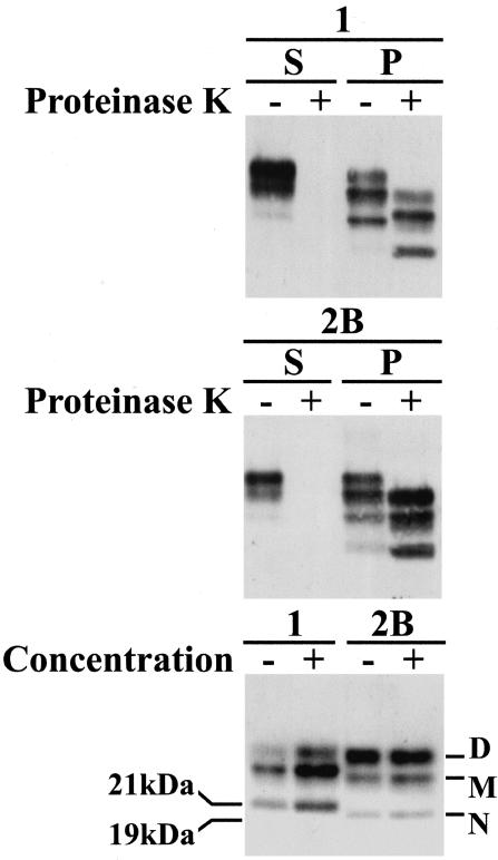
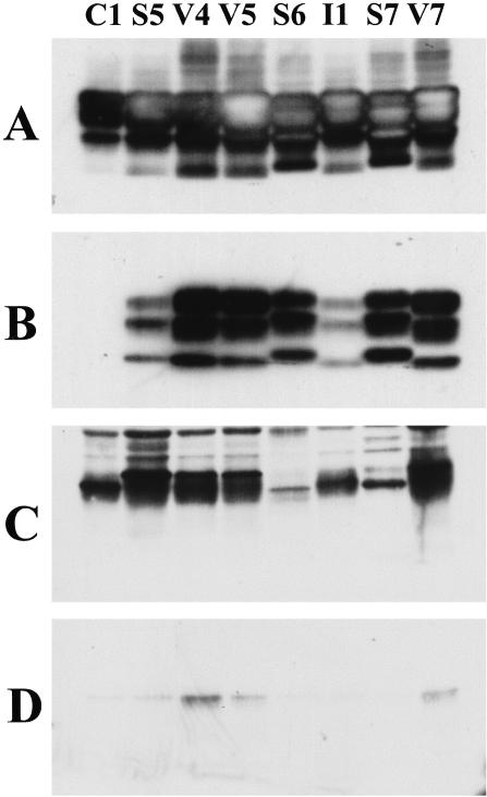
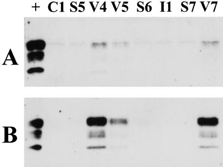
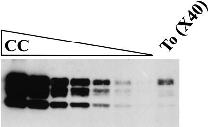
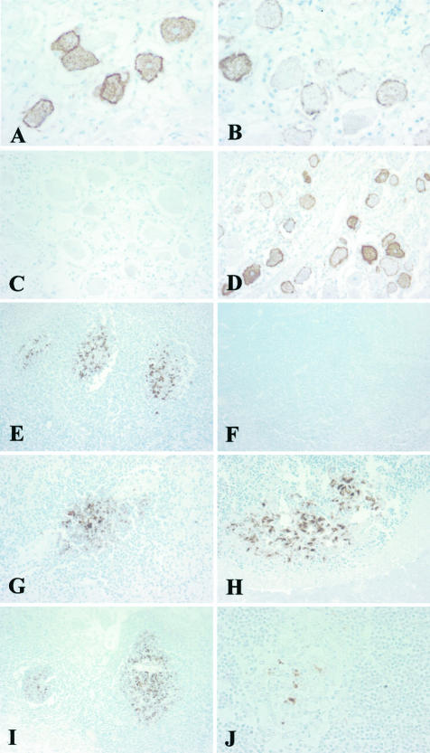
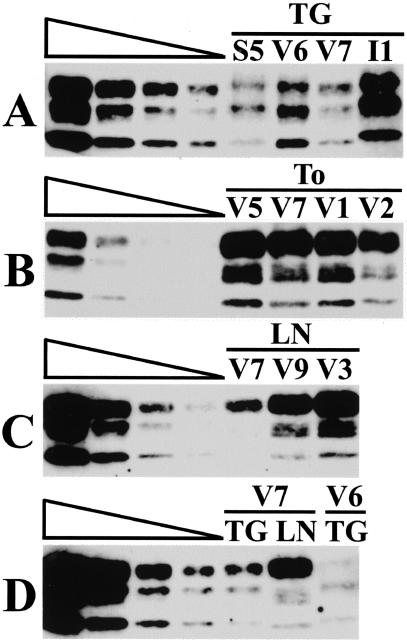
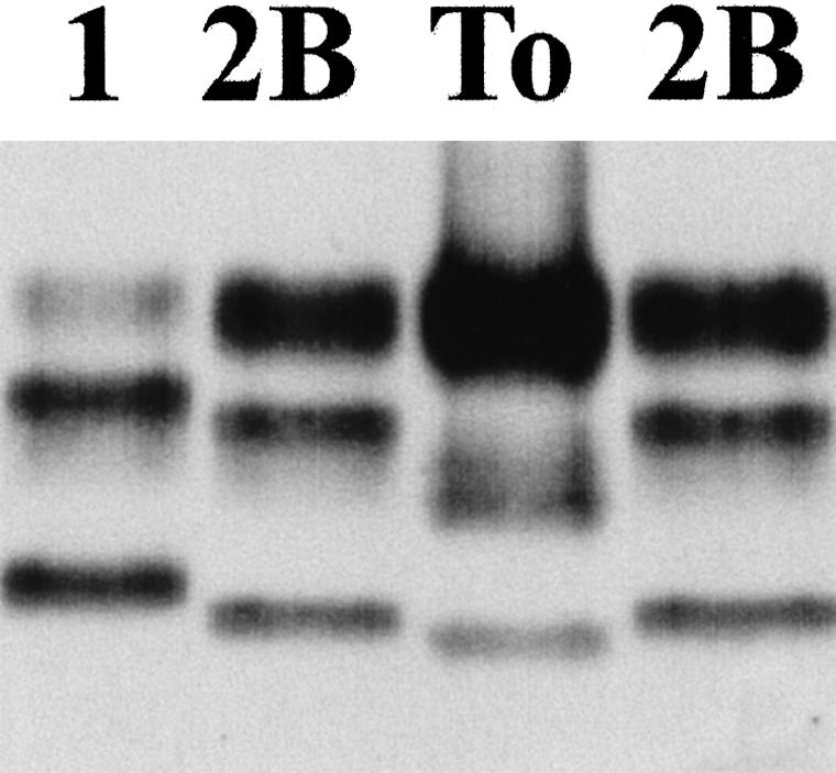
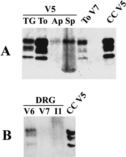
Human prion diseases are rare fatal neurodegenerative conditions that occur as acquired, familial, or idiopathic disorders. A key event in their pathogenesis is the accumulation of an altered form of the prion protein, termed PrP(Sc), in the central nervous system. A novel acquired human prion disease, variant Creutzfeldt-Jakob disease, is thought to result from oral exposure to the bovine spongiform encephalopathy agent. This disease differs from other human prion diseases in its neurological, neuropathological, and biochemical phenotype. We have used immunohistochemistry and Western blot techniques to analyze the tissue distribution and biochemical properties of PrP(Sc) in peripheral tissues in a unique series of nine cases of variant Creutzfeldt-Jakob disease. We have compared this with the distribution and biochemical forms found in all of the major subtypes of sporadic Creutzfeldt-Jakob disease and in a case of iatrogenic Creutzfeldt-Jakob disease associated with growth hormone therapy. The results show that involvement of the lymphoreticular system is a defining feature of variant Creutzfeldt-Jakob disease, but that the biochemical isoform of PrP(Sc) found is influenced by the cell type in which it accumulates.


Figures

Figure 1
Figure 1
Western blot analysis of standard frontal cortex extracts from sporadic CJD type 1 (1) and variant CJD type 2B (2B). Samples were separated by centrifugation at 21,000 × g into supernatant (S) or pellet (P) fraction. These two fractions were then analyzed before (−) or after (+) digestion with proteinase K as shown in the top and middle panel. In a separate experiment, the same extracts were digested with proteinase K and an aliquot diluted by a factor of 40, collected by centrifugation, and resuspended in the original volume. Analysis of equivalent amounts of the original (−) and the diluted/concentrated samples (+) are shown at the bottom. The 21-kd nonglycosylated fragment diagnostic of type 1 PrPres and the 19-kd nonglycosylated fragment diagnostic of type 2 PrPres are marked in the bottom panel. The positions of the three glycoforms; diglycosylated (D), monoglycosylated (M), and nonglycosylated (N) are also indicated.
Figure 2
Figure 2
Western blot analysis of frontal cortex (A and B) and tonsil (C and D) analyzed with (B and D) or without (A and C) previous digestion with proteinase K from cases C1 (Lewy body dementia), S5, S6, S7 (sCJD) V4, V5, V7 (vCJD), and I1 (iCJD). All blots were loaded with an equivalent amount of tissue extract and processed together but the exposure time of A and B (frontal cortex) was 1/36th of that for C and D (tonsil).
Figure 3
Figure 3
Western blot analysis of PrPres in tonsil samples from cases C1 (Lewy body dementia), S5, S6, S7 (sCJD) V4, V5, V7 (vCJD), and I1 (iCJD) before (A) and after (B) centrifugal enrichment for PrPres from an eightfold larger volume of extract. +, Indicates a lane loaded with a one-tenth volume of the frontal cortex from vCJD case V7. Both blots were processed together and exposed for the same length of time.
Figure 4
Figure 4
Western blot analysis of a dilution series of cerebral cortex (CC) and concentrated tonsil (To) extract from a case of vCJD (V7). The serial dilution was by a factor of two, starting with a loading of 5 μl of a 10% extract of frontal cortex in the first lane. The lane of tonsil represents 200 μl of a 10% extract enriched (×40) by centrifugal concentration for PrPres.
Figure 5
Figure 5
Immunohistochemical analysis of PrP showing intense staining of dorsal root ganglion cells in variant CJD, case V2 (A); staining of occasional dorsal root ganglion cells in sporadic CJD, case S1 (B); no reactivity in the dorsal root ganglion in another sporadic CJD case of different genotype, case S3 (C); variable staining of ganglion cells in the trigeminal ganglion in sporadic CJD, case S4 (D); intense immunoreactivity within germinal centers in the tonsil in variant CJD, case V3 (E); no reactivity in the tonsil in sporadic CJD, case S1 (F); staining of germinal centers in variant CJD in the spleen, appendix, and an ileal Peyer’s patch, respectively, case V2 (G–I); patchy staining within a small germinal center in the thymus in variant CJD, case V4 (J). The monoclonal antibody KG9 was used in all images. Original magnifications: ×400 (A, B, J); ×200 (C, D, G, H); ×100 (E, F, I).
Figure 6
Figure 6
Western blot analysis of concentrated extracts of trigeminal ganglion (TG), tonsil (To), and cervical lymph node (LN). The cases are identified as S (sporadic CJD), V (variant CJD), and I (iatrogenic CJD) and the case number using the codes shown in Table 1. A twofold dilution series of frontal cortex from case V5 is shown extending left to right (undiluted, 1:2, 1:4, 1:8). The signals from the tonsil samples shown in B are not comparable to any lanes of the corresponding frontal cortex dilution series and the estimation of PrPres concentration for these particular samples was made from concentrates using less starting tonsil extract.
Figure 7
Figure 7
Western blot analysis of PrPres in a concentrated extract made from tonsil biopsy tissue (To). A standard type 1 PrPres isotype from sporadic CJD frontal cortex (1) and a standard type 2B PrPres isotype from variant CJD frontal cortex (2B) are also shown.
Figure 8
Figure 8
Western blot analysis of PrPres in (A) concentrated extracts of trigeminal ganglion (TG), tonsil (To), appendix (Ap), and spleen (Sp) from a case of vCJD (V5) and in (B) concentrated extracts of dorsal root ganglion (DRG) from cases of vCJD and iCJD (V6, V7, I1). Concentrated tonsil from a case vCJD (To V7) and unconcentrated frontal cortex from a case of vCJD (CC V5) are included as positive controls.

References

    1. Prusiner SB. Prions. Proc Natl Acad Sci USA. 1998;95:13363–13383. - PMC - PubMed
    1. Brown P, Preece M, Brandel J-P, Sato T, McShane L, Zerr I, Fletcher A, Will RG, Pocchiari M, Cashman NR, d‘Aignaux JH, Cervenakova L, Fradkin J, Schonberger LB, Collins SJ. Iatrogenic Creutzfeldt-Jakob disease at the millennium. Neurology. 2000;55:1075–1081. - PubMed
    1. Deslys JP, Marce D, Dormont D. Similar genetic susceptibility in iatrogenic and sporadic Creutzfeldt-Jakob disease. J Gen Virol. 1994;1:23–27. - PubMed
    1. Monari L, Chen SG, Brown P, Parchi P, Petersen RB, Mikol J, Gray F, Cortelli P, Montagna P, Ghetti B, Goldfarb LG, Gajdusek DC, Lugaresi E, Gambetti P, Autilio-Gambetti L. Fatal familial insomnia and familial Creutzfeldt-Jakob disease: different prion proteins determined by a DNA polymorphism. Proc Natl Acad Sci USA. 1994;91:2839–2842. - PMC - PubMed
    1. Parchi P, Castellani R, Capellari S, Ghetti B, Young K, Chen SG, Farlow M, Dickson DW, Sima AAF, Trojanowski JQ, Petersen RB, Gambetti P. Molecular basis of phenotypic variability in sporadic Creutzfeldt-Jakob disease. Ann Neurol. 1996;39:767–78. - PubMed

Publication types