Comparative Study
. 2004 Jan;164(1):155-66.
doi: 10.1016/s0002-9440(10)63106-9.

Expression of the small heat-shock protein alphaB-crystallin in tauopathies with glial pathology

Affiliations
Comparative Study

Expression of the small heat-shock protein alphaB-crystallin in tauopathies with glial pathology

Deepa V Dabir et al. Am J Pathol. 2004 Jan.

Abstract

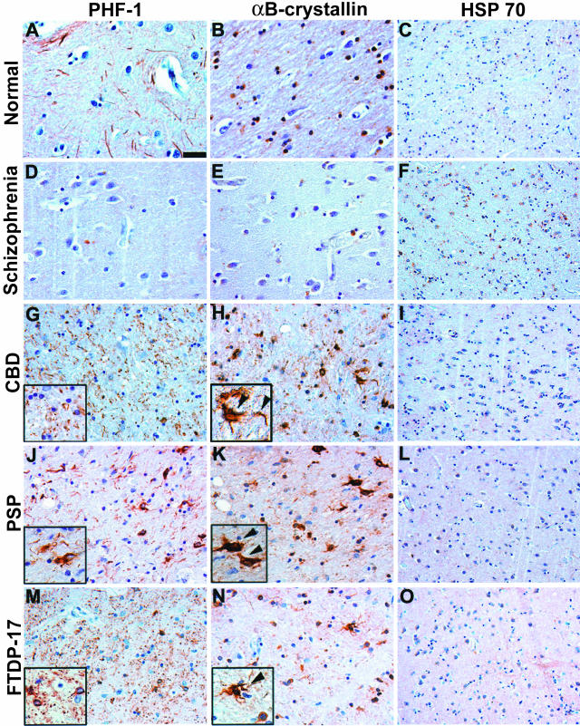
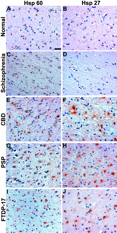
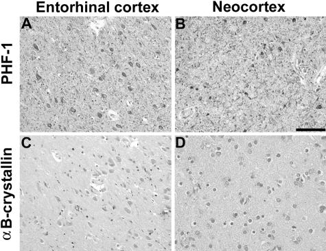
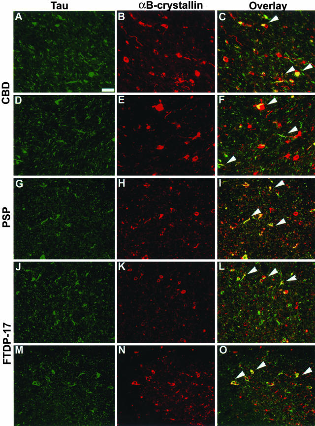
Intracellular accumulations of filamentous material composed of tau proteins are defining features of sporadic and familial neurodegenerative disorders termed "tauopathies." In Alzheimer's disease, the most common tauopathy, tau pathology is predominantly localized within neurons; however, robust glial pathology occurs in other tauopathies. Although the pathogenesis of tauopathies remains primarily unknown, molecular chaperones such as heat-shock proteins (HSPs) are implicated in these tau disorders as well as other neurodegenerative diseases characterized by the accumulation of insoluble protein aggregates such as alpha-synuclein in Parkinson's disease and polyglutamine in Huntington's disease. We analyzed a variety of tauopathies with antibodies to a panel of HSPs to determine their role in the pathogenesis of these disorders. Although HSPs are not found in neuronal tau inclusions, we demonstrate increased expression of the small HSP alphaB-crystallin in glial inclusions of both sporadic and familial tauopathies. alphaB-crystallin was observed in a subset of astrocytic and oligodendrocytic tau inclusions as well as the neuropil thread pathology in cellular processes, but the co-expression of alphaB-crystallin with tau inclusions was relatively specific to tauopathies with extensive glial pathology. Thus, increased alphaB-crystallin expression in glial tau inclusions may represent a response by glia to the accumulation of misfolded or aggregated tau protein that is linked to the pathogenesis of the glial pathology and distinct from mechanisms underlying neuronal tau pathology in neurodegenerative disease.


Figures

Figure 1
Figure 1
Expression of αBC in subcortical white matter of patients with tauopathies. Adjacent sections of mid-frontal [normal, schizophrenia (patient 2), PSP (patient 2), and FTDP-17 (patient 1)] or temporal cortex [CBD (patient 5)] were immunostained with PHF1 (A, D, G, J, M), αBC (B, E, H, K, N), or HSP70 (C, F, I, L, O). A, D, G, J, and M: Robust tau pathology including threads and coiled bodies was observed in the subcortical white matter of CBD and FTDP-17 brains. The PSP brain showed mild tau pathology whereas no pathology was detected in schizophrenia or normal control brains. Insets in G and M show high-power magnification of characteristic glial pathology in CBD and FTDP-17 including coiled bodies and neuropil threads. B, E, H, K, and N: Adjacent sections show robust αBC staining in regions with abundant tau pathology of CBD and FTDP-17 brains. Insets in H and N show αBC staining in CBD and FTDP-17 that resembles coiled bodies (arrowheads). PSP, which showed mild tau pathology, also showed mild αBC immunoreactivity (arrowheads). In contrast, the control and schizophrenia brains show normal levels of αBC staining. C, F, I, L, and O: No increase in the iHSP70 immunoreactivity was observed in disease relative to control brains. Scale bar, 100 μm (A, B, D, E, G, H, J, K, M, N); 200 μm (C, F, I, L, O).
Figure 2
Figure 2
Expression of αBC in the basal ganglia of patients with tauopathies. Adjacent sections of basal ganglia were immunostained with PHF1 (A, D, G, J, M), αBC (B, E, H, K, N), and HSP70 (C, F, I, L, O). A, D, G, J, and M: Robust tau pathology including threads and coiled bodies was observed in the globus pallidus and to a lesser extent, striatum in CBD (patient 2), PSP (patient 6), and FTDP-17 (patient 1) brains. No tau pathology was detected in the brains from schizophrenia (patient 2) or normal controls. Insets in G, J, and M show high-power magnifications of characteristic glial pathology in CBD, PSP, and FTDP-17 including coiled bodies and neuropil threads. B, E, H, K, and N: Similar to the neocortex, adjacent sections show robust αBC staining in sections with abundant tau pathology (H, K, N). Insets show αBC staining that resembles coiled bodies (arrowheads, H and K) in CBD and PSP or astrocytic inclusions (N) in FTDP-17. In contrast, the normal and schizophrenia brains show baseline levels of αBC staining. C, F, I, L, and O: No increase in iHSP70 immunoreactivity was detected in the basal ganglia of tauopathies. Scale bar, 100 μm (A, B, D, E, G, H, J, K, M, N); 200 μm (C, F, I, L, O).
Figure 3
Figure 3
αBC expression is increased in affected brain regions of patients with tauopathies. The mid-frontal and occipital cortex (gray and white matter) and basal ganglia (globus pallidus and caudate/putamen) from five CBD, six PSP, two FTDP-17, and six schizophrenia (SCH) were analyzed by IHC for tau and αBC. Immunoreactivity was graded semiquantitatively as 0 (absent), 1 (mild), 2 (moderate), or 3 (severe). Bars in the graph represent mean tau pathology (open bars) and αBC immunoreactivity (solid bars). Error bars indicate SD of the mean. *, Denotes regions with a mean value of zero.
Figure 4
Figure 4
HSP60 and HSP27 immunoreactivity in tauopathies. Adjacent sections of neocortex as shown in Figure 1 were immunostained with HSP60 (A, C, E, G, I) and HSP27 (B, D, F, H, J). A, C, E, G, and I: HSP60 immunoreactivity was detected in reactive astrocytes in the neocortex of CBD (patient 5), PSP (patient 6), and FTDP-17 (patient 1) patients. Similar HSP60 staining was also observed in the basal ganglia (data not shown). B, D, F, H, and J: HSP27 staining was increased in CBD, PSP, and FTDP-17 brains predominantly in glial cells in the neocortex. The normal and schizophrenia brains show baseline levels of HSP60 (A, C) and HSP27 (B, D) staining. Scale bar, 100 μm.
Figure 5
Figure 5
The increase in αBC expression is relatively specific to glial pathology. Adjacent sections from AD entorhinal (A, C) and frontal cortex (B, D) were immunostained with PHF1 (A, B) and αBC (C, D). A and B: The AD brain sections show robust tau pathology in entorhinal cortex and neocortex. C and D: In contrast, immunostaining for αBC is similar to that observed in control and schizophrenia brains. Similar results were detected in the brains of six patients pathologically diagnosed as AD. In contrast, reactive astrocytes in the subcortical white matter were focally positive for αBC, as described previously (data not shown). Scale bar, 100 μm.
Figure 6
Figure 6
Co-localization of αBC with tau-immunoreactive inclusions. Adjacent sections from affected regions were double stained for tau (green; A, D, G, J, M) and αBC (red; B, E, H, K, N). Merged images are depicted in C, F, I, L, and O. αBC co-localized with a subset of glial tau pathology in the neocortex of CBD (A–C, patient 5) and FTDP-17 (J–L, patient 1). Similar co-localization was also observed in the basal ganglia of CBD (D–F, patient 2), PSP (G–I, patient 2) and FTDP-17 (M–O, patient 1). Arrowheads indicate some of the inclusions in which co-localization was observed. Scale bar, 100 μm.
Figure 7
Figure 7
Biochemical analysis of tauopathy brains demonstrate increased insoluble αBC in affected brain regions. Sequential tau extractions from frontal gray (A) and white (B) matter, globus pallidus (C), and caudate/putamen (D) from PSP, CBD, schizophrenia (Sch), and normal (N) brains were performed as described. Soluble (HS-TBS) and insoluble (2% SDS and 70% FA) fractions were generated and analyzed by SDS-polyacrylamide gel electrophoresis followed by immunoblotting for tau using a cocktail of Tau14 and Tau 46 (brackets, top) and mAbs specific for αBC (arrow, bottom). The soluble extracts showed robust tau expression and variable levels of αBC in both affected and control brains. In contrast, there were increased amounts of insoluble αBC (FA > SDS) in brains from tauopathy patients that paralleled the elevated levels of insoluble tau (see Figure 8).
Figure 8
Figure 8
Quantitative Western blot analyses of insoluble tau shows a marked increase in insoluble αBC in affected brain regions of some tauopathies. Quantitative Western blots of insoluble SDS (open bars) and FA (solid bars) αBC fractions from frontal gray and white matter, globus pallidus, and caudate/putamen were performed using I125-labeled secondary antibodies. SDS- and FA-insoluble fractions from schizophrenia (SCH), CBD, and PSP cases were individually compared with the corresponding fractions from normal (N) cases. Graph represents average fold increase in αBC expression compared to normal brains. There is a marked increase in insoluble αBC detected in FA, and to a lesser extent in SDS extracts. Error bars represent SD of the mean.

References

    1. Lee VM-Y, Goedert M, Trojanowski JQ. Neurodegenerative tauopathies. Ann Rev Neurosci. 2001;24:1121–1159. - PubMed
    1. Cleveland DW, Hwo SY, Kirschner MW. Purification of tau, a microtubule-associated protein that induces assembly of microtubules from purified tubulin. J Mol Biol. 1977;116:207–225. - PubMed
    1. Buée L, Bussière T, Buée-Scherrer V, Delacourte A, Hof PR. Tau protein isoforms, phosphorylation and role in neurodegenerative disorders. Brain Res Rev. 2000;33:1–36. - PubMed
    1. Komori T. Tau-positive glial inclusions in progressive supranuclear palsy, corticobasal degeneration and Pick’s disease. Brain Pathol. 1999;9:663–679. - PMC - PubMed
    1. Shin RW, Iwaki T, Kitamoto T, Tateishi J. Hydrated autoclave pretreatment enhances tau immunoreactivity in formalin-fixed normal and Alzheimer’s disease brain tissues. Lab Invest. 1991;64:693–702. - PubMed

Publication types

LinkOut - more resources