Comparative Study
. 2004 Jan;164(1):253-62.
doi: 10.1016/S0002-9440(10)63115-X.

S100A4/Mts1 produces murine pulmonary artery changes resembling plexogenic arteriopathy and is increased in human plexogenic arteriopathy

Affiliations
Comparative Study

S100A4/Mts1 produces murine pulmonary artery changes resembling plexogenic arteriopathy and is increased in human plexogenic arteriopathy

Steven Greenway et al. Am J Pathol. 2004 Jan.

Abstract

S100A4/Mts1 confers a metastatic phenotype in tumor cells and may also be related to resistance to apoptosis and angiogenesis. Approximately 5% of transgenic mice overexpressing S100A4/Mts1 develop pulmonary arterial changes resembling human plexogenic arteriopathy with intimal hyperplasia leading to occlusion of the arterial lumen. To assess the pathophysiological significance of this observation, immunohistochemistry was applied to quantitatively analyze S100A4/Mts1 expression in pulmonary arteries in surgical lung biopsies from children with pulmonary hypertension secondary to congenital heart disease. S100A4/Mts1 was not detected in pulmonary arteries with low-grade hypertensive lesions but was expressed in smooth muscle cells of lesions showing neointimal formation and with increased intensity in vessels with an occlusive neointima and plexiform lesions. Putative downstream targets of S100A4/Mts1 include Bax, which is pro-apoptotic, and the pro-angiogenic vascular endothelial growth factor (VEGF). The increase in S100A4/Mts1 expression precedes heightened expression of Bax in progressively severe neointimal lesions but in non-S100A4/Mts1-expressing cells. VEGF immunoreactivity did not correlate with severity of disease. The relationship of increased S100A4/Mts1 to pathologically similar lesions in the transgenic mice and patients occurs despite differences in localization (endothelial versus smooth muscle cells).


Figures
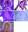
Figure 1
Figure 1
Representative photomicrographs of mouse lung tissue. A: Movat pentachrome staining of lung tissue from a transgenic mouse engineered to overexpress S100A4/Mts1 showing an occlusive neointimal lesion consistent with plexogenic arteriopathy. B: A representative Movat pentachrome-stained pulmonary artery from an age-matched, non-transgenic mouse shows a normal thin walled vessel with a patent lumen. C: Lung tissue section stained with hematoxylin and eosin from a transgenic mouse engineered to overexpress S100A4/Mts1, demonstrates increased perivascular inflammatory cells. D: Immunoperoxidase staining with an antibody to von Willebrand factor in a vessel with extensive neointimal formation from a mouse engineered to overexpress S100A4/Mts1 shows that the cells lining the lumen are endothelial. E: Plexogenic-like lesion in a S100A4/Mts1 transgenic mouse stained with α-actin shows numerous neointimal smooth muscle cells. Original magnification, ×40. Bar, 50 μm.
Figure 2
Figure 2
Representative photomicrographs of lung tissue from diseased, transgenic mice engineered to overexpress S100A4/Mts1. A: Immunoperoxidase staining for S100A4/Mts1 in pulmonary arteries with occlusive neointimal lesions shows expression in endothelial cells and in some periadventitial cells which appear by morphology to be inflammatory. B: Immunoperoxidase staining for Bax is present in some of the periadventitial cells surrounding an artery with an occlusive neointimal lesion as indicated by the arrows. C: Pulmonary artery stained for PCNA. Arrows indicate an occasional positive cell within the neointima of the lesion but most are present in the periadventitia. D: Staining for VEGF reveals the absence of positive cells within the artery. Original magnification, ×40.
Figure 3
Figure 3
Representative photomicrographs of human lung biopsy tissue after immunoperoxidase staining for S100A4/Mts1. A: Vessel from patient graded 0-IB showing normal pulmonary artery with no immunodetectable S100A4/Mts1. B: Vessel showing a typical grade IB lesion with severe medial hypertrophy but without immunoreactivity for S100A4/Mts1. C: An artery from a patient with grade IVC disease with occlusive neointimal proliferation and strong positive staining for S100A4/Mts1 particularly in the intima compared to the media of the vessel. S100A4/Mts1 was not detected in all cells and appears to be localized in a subpopulation of intimal cells. D: A plexogenic lesion from a patient with grade IVC disease with staining of the smooth muscle cells and sparing of the endothelial cells (arrows). Immunoreactivity for S100A4/Mts1 was present in the lung parenchyma at a similar level in all grades of pulmonary vascular disease. Original magnification, ×40.
Figure 4
Figure 4
The expression of S100A4/Mts1 in pulmonary arteries from children with congenital heart disease increases as the severity of pulmonary hypertension increases. The intensity of immunohistochemical staining for S100A4/Mts1 in lung biopsy tissue was quantified as described in Materials and Methods. Groups II, III, and IV are significantly different from group I (P value <0.05 compared to group 1). Graphs represent mean values ± SEM.
Figure 5
Figure 5
Representative photomicrographs of human lung biopsy tissue. A: Note the relative absence of Bax in a pulmonary artery graded 0–1B. B: A grade IVC vessel stained with von Willebrand factor to demonstrate endothelial cells within the plexogenic lesion. C and D: Serial sections comparing the distribution of Bax (C) and S100A4/Mts1 (D) expression in grade IVC disease. Bax expression is noted in the endothelial cells and non-S100A4/Mts1-expressing medial smooth muscle cells (arrows). S100A4/Mts1 expression occurs primarily in the intima. Original magnification, ×40.
Figure 6
Figure 6
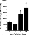
There is increased expression of Bax in the pulmonary arteries from children with pulmonary hypertension secondary to congenital heart disease in severe pulmonary hypertension (grades III and IV). The intensity of immunohistochemical staining for Bax in lung biopsy tissue was quantified as described in Materials and Methods. Groups III and IV are significantly different from group I (P value <0.05). Graphs represent mean values ± SEM.
Figure 7
Figure 7
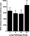
Expression of VEGF in the pulmonary arteries from children with pulmonary hypertension secondary to congenital heart disease remains relatively constant despite increasing severity of disease. The intensity of immunohistochemical staining for VEGF in lung biopsy tissue was quantified as described in Materials and Methods. Graphs represent mean values ± SEM.

References

    1. Rabinovitch M. Diseases of the pulmonary vasculature. Topol EJ, editor. Philadelphia: Lippincott-Raven,; Comprehensive Cardiovascular Medicine. 1998:pp 3001–3029.
    1. Rabinovitch M, Haworth SG, Castaneda AR, Nadas AS, Reid LM. Lung biopsy in congenital heart disease: a morphometric approach to pulmonary vascular disease. Circulation. 1978;58:1107–1122. - PubMed
    1. Heath D, Edwards JE. The pathology of hypertensive pulmonary vascular disease. Circulation. 1958;18:533–547. - PubMed
    1. Rabinovitch M. Pathobiology of pulmonary hypertension: extracellular matrix. Clin Chest Med. 2001;22:433–449. - PubMed
    1. Geraci M, Gao B, Shepherd D, Moore M, Westcott J, Fagan K, Alger L, Tuder R, Voelkel N. Pulmonary prostacyclin synthase overexpression in transgenic mice protects against development of hypoxic pulmonary hypertension. J Clin Invest. 1999;103:1509–1515. - PMC - PubMed

Publication types

MeSH terms