. 2004 Jan 6;101(1):296-301.
doi: 10.1073/pnas.2434651100. Epub 2003 Dec 26.

Reversing established sepsis with antagonists of endogenous high-mobility group box 1

Affiliations

Reversing established sepsis with antagonists of endogenous high-mobility group box 1

Huan Yang et al. Proc Natl Acad Sci U S A. .

Abstract

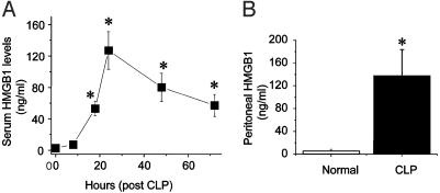
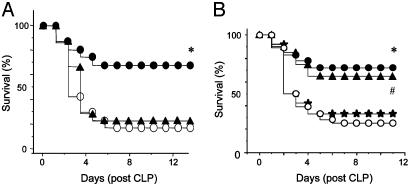
Despite significant advances in intensive care therapy and antibiotics, severe sepsis accounts for 9% of all deaths in the United States annually. The pathological sequelae of sepsis are characterized by a systemic inflammatory response, but experimental therapeutics that target specific early inflammatory mediators [tumor necrosis factor (TNF) and IL-1beta] have not proven efficacious in the clinic. We recently identified high mobility group box 1 (HMGB1) as a late mediator of endotoxin-induced lethality that exhibits significantly delayed kinetics relative to TNF and IL-1beta. Here, we report that serum HMGB1 levels are increased significantly in a standardized model of murine sepsis, beginning 18 h after surgical induction of peritonitis. Specific inhibition of HMGB1 activity [with either anti-HMGB1 antibody (600 microg per mouse) or the DNA-binding A box (600 microg per mouse)] beginning as late as 24 h after surgical induction of peritonitis significantly increased survival (nonimmune IgG-treated controls = 28% vs. anti-HMGB1 antibody group = 72%, P < 0.03; GST control protein = 28% vs. A box = 68%, P < 0.03). Animals treated with either HMGB1 antagonist were protected against the development of organ injury, as evidenced by improved levels of serum creatinine and blood urea nitrogen. These observations demonstrate that specific inhibition of endogenous HMGB1 therapeutically reverses lethality of established sepsis indicating that HMGB1 inhibitors can be administered in a clinically relevant time frame.


Figures

Fig. 1.
Fig. 1.
Serum and peritoneal HMGB1 levels are elevated in septic mice. BALB/c mice (male, 20-25 g) were subjected to CLP, and sera were collected at various time points and assayed for HMGB1 levels as described (11). (A) Serum HMGB1 was not detectable until 18 h after surgery and then remained elevated from 24 to 72 h after surgery. Data are shown as mean ± SEM (n = 3-6 mice per time point). *, P < 0.05 vs. control as tested by one-way ANOVA followed by the least-significant difference test. (B) Peritoneal exudate fluid was collected 48 h after cecal perforation or sham surgery (normal), and HMGB1 was measured. Data are mean ± SEM of 7 and 11 mice per group. *, P < 0.05 vs. normal as tested by Student's t test.
Fig. 2.
Fig. 2.
Anti-HMGB1 antibodies specifically inhibit HMGB1-induced cytokine release. Murine macrophage-like RAW 264.7 cells were stimulated with HMGB1, IL-1β, or TNF as indicated for 16 h in serum-free Opti-MEM medium in the absence or presence of purified rabbit anti-HMGB1 IgG antibodies or nonimmune rabbit IgG (as control). Conditioned media were collected and assayed for TNF and IL-6 levels by use of ELISA kits. Data represent mean ± SEM of three to six independent experiments, each done in duplicate. *, P < 0.05 vs. control; **, P < 0.05 vs. HMGB1 alone.
Fig. 3.
Fig. 3.
Anti-HMGB1 antibodies protect against lethality caused by cecal perforation. (A) BALB/c mice were randomly grouped (18-25 mice per group) and subjected to CLP. Mice received neutralizing IgG-purified (or, in some experiments, affinity-purified) anti-HMGB1 antibodies twice daily for 3 days at 60 or 600 μg per mouse (i.p.) beginning 24 h after surgery. Irrelevant nonimmune rabbit IgG was used as control. *, P < 0.03 vs. control, as tested by two-tailed Fisher's exact test. •, anti-HMGB1 antibody, 600 μg per mouse (24-h delay); ▴, anti-HMGB1 antibody, 60 μg per mouse (24-h delay); ○, IgG control. (B) Anti-HMGB1 antibody treatment (600 μg per mouse) was initiated at 12, 24, or 36 h after surgery, twice a day, and continued for 3 days. * and #, P < 0.05 vs. IgG control group. ▴, anti-HMGB1 antibody (12-h delay); •, anti-HMGB1 antibody (24-h delay); ★, anti-HMGB1 antibody (36-h delay); ○, IgG control.
Fig. 4.
Fig. 4.
Schematic representation of HMGB1 constructs and their activity in TNF stimulation. Subconfluent cultures of RAW 264.7 cells were incubated with various HMGB1 mutant proteins (1 μg/ml) for 8 h. The conditioned medium was assayed for TNF by a standard murine fibroblast L929 (ATCC) cytotoxicity bioassay (20). Wild-type HMGB1, carboxyl-truncated, and B box significantly stimulated TNF release (*, P < 0.05 vs. GST control, one-way ANOVA followed by the least-significant difference test), but A box stimulation of TNF was not statistically significant. Full-length HMGB1 = 216 aa; carboxyl-truncated mutant corresponds to amino acids 1-182 of HMGB1; B box corresponds to amino acids 88-162 of HMGB1; and A box corresponds to amino acids 1-85 of HMGB1. n = 6-10 separate experiments.
Fig. 5.
Fig. 5.
A box antagonizes HMGB1 in vitro. Subconfluent RAW 264.7 cells in 24-well dishes were treated with HMGB1 or IL-1β and various concentrations of A box (as indicated) for 16 h in Opti-MEM I medium in the presence of polymyxin B. The TNF- or IL-1β-stimulating activity was expressed as percent of maximal response, and inhibition by A box was expressed as percent of HMGB1 or IL-1β alone. (A) HMGB1 (1 μg/ml) induced the release of 13,344 ± 2,335 pg of TNF per ml. A box dose-dependently inhibited HMGB1-induced TNF release with an apparent EC50 of 7.5 μg/ml. (B) Similarly, HMGB1-induced IL-1β was dose-dependently inhibited by the addition of A box. (C) In contrast, A box did not have any significant effect on IL-1β-induced TNF release. TNF release stimulated by IL-1β (100 ng/ml) was 640 ± 160 pg of TNF per ml. (D) RAW 267.4 cells were stimulated with LPS and A box, B box, or GST vector, or LPS at the concentration indicated for 16 h, and IL-10 released in the conditioned medium was measured with an ELISA kit. Data represent three or four separate experiments.
Fig. 6.
Fig. 6.
A box protects against lethality caused by LPS or cecal perforation in mice. (A) BALB/c mice were randomly grouped (18 mice per group) and challenged with an LD75 dose of LPS (15 mg/kg of body weight, i.p.). A box or GST control was administered at 600 μg per mouse (in 300 μl of sterile PBS) either immediately or 2 h after LPS injection. Two additional doses were given (i.p.) at 12 and 24 h after the first treatment. * and #, P < 0.05 vs. GST control group as determined by two-tailed Fisher's exact test. •, A box (0-h delay); ▴, A box (2-h delay); ○, GST control. (B) BALB/c mice (15-25 mice per group) were subjected to CLP. At 24 h after the operation, mice were given recombinant HMGB1 A box or GST control (60 or 600 μg per mouse per injection, i.p.) twice daily for 3 days, beginning 24 h after surgery. Animal survival was monitored for up to 2 weeks to ensure that no late death occurred. *, P < 0.03 vs. GST control as tested by two-tailed Fisher's exact test. ▴, A box 60 μg per mouse (24-h delay); •, A box 600 μg per mouse (24-h delay); ○, GST control.

References

    1. Munford, R. S. & Pugin, J. (2001) Am. J. Respir. Crit. Care Med. 163, 316-321. - PubMed
    1. Angus, D. & Wax, R. S. (2001) Crit. Care Med. 29, Suppl. 7, S109-S116. - PubMed
    1. Friedman, G., Silva, E. & Vincent, J. L. (1998) Crit. Care Med. 26, 2078-2086. - PubMed
    1. Tracey, K. J., Beutler, B., Lowry, S. F., Merryweather, J., Wolpe, S., Milsark, I. W., Hariri, R. J., Fahey, T. J., III, Zentelia, A., Albert, J. D., et al. (1986) Science 234, 470-474. - PubMed
    1. Tracey, K. J., Fong, Y., Hesse, D. G., Manogue, K. R., Lee, A. T., Kuo, G. C., Lowry, S. F. & Cerami, A. (1987) Nature 330, 662-664. - PubMed

Publication types