. 2004 Jan 1;265(1):181-95.
doi: 10.1016/j.ydbio.2003.09.019.

HB-EGF directs stromal cell polyploidy and decidualization via cyclin D3 during implantation

Affiliations

HB-EGF directs stromal cell polyploidy and decidualization via cyclin D3 during implantation

Yi Tan et al. Dev Biol. .

Abstract

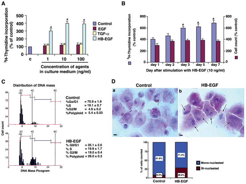
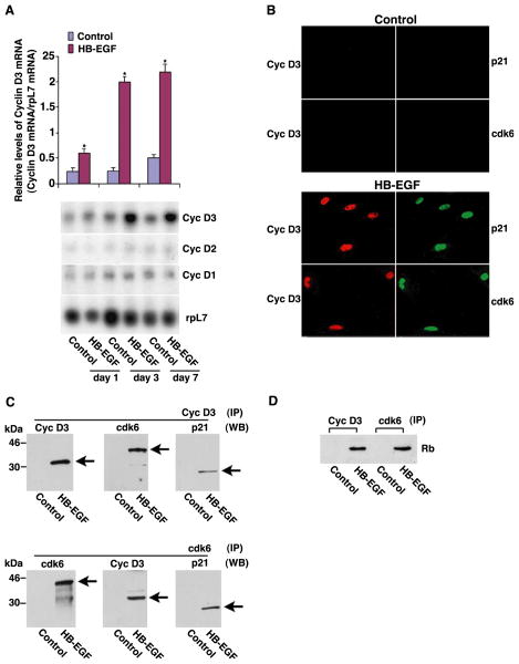
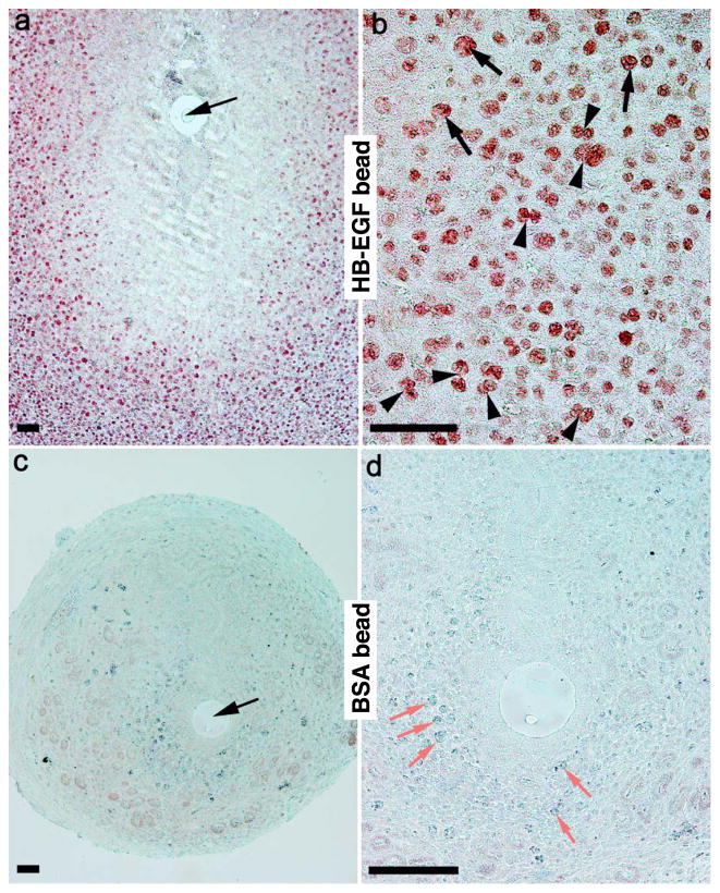
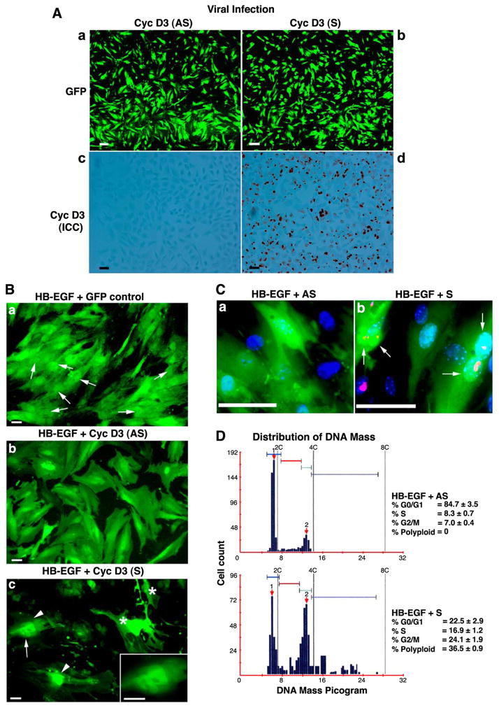
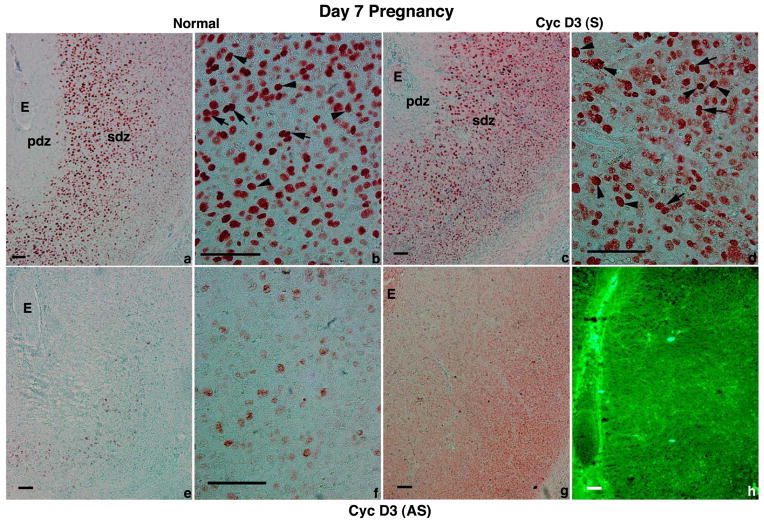
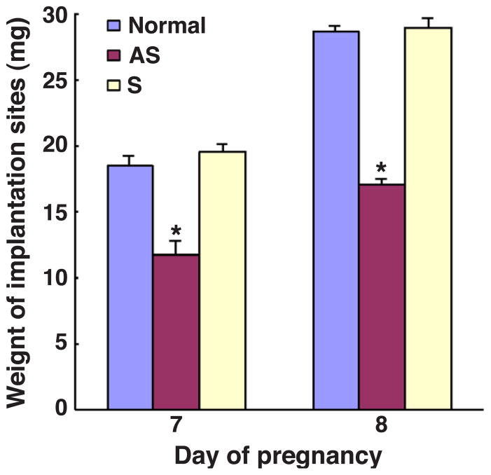
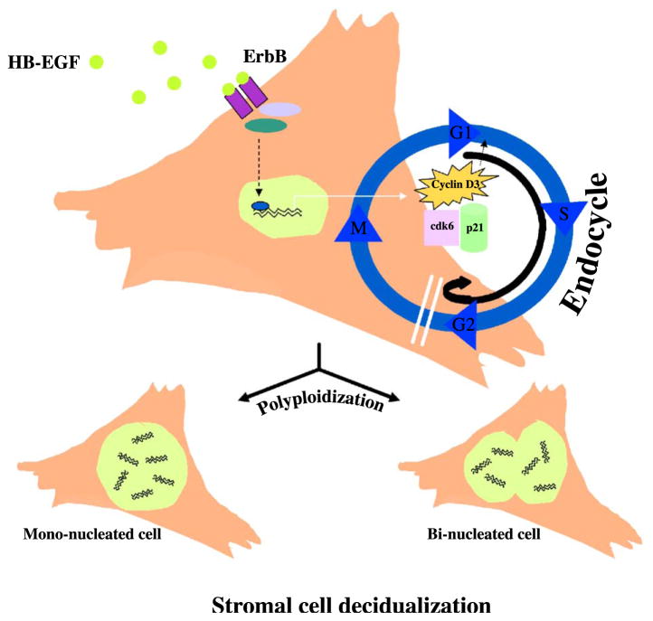
Stromal cell polyploidy is a unique phenomenon that occurs during uterine decidualization following embryo implantation, although the developmental mechanism still remains elusive. The general consensus is that the aberrant expression and altered functional activity of cell cycle regulatory molecules at two particular checkpoints G1 to S and G2 to M in the cell cycle play an important role in the development of cellular polyploidy. Despite the compelling evidence of intrinsic cell cycle alteration, it has been implicated that the development of cellular polyploidy may be controlled by specific actions of extracellular growth regulators. Here we show a novel role for heparin-binding EGF-like growth factor (HB-EGF) in the developmental process of stromal cell polyploidy in mice. HB-EGF, which is one of the earliest known molecular mediators of implantation in mice and humans, promotes stromal cell polyploidy via upregulation of cyclin D3. Adenoviral delivery of antisense cyclin D3 attenuates cyclin D3 expression and abrogates HB-EGF-induced stromal cell polyploidy in vitro and in vivo. Collectively, the results demonstrate that the regulation of stromal cell polyploidy and decidualization induced by HB-EGF depend on cyclin D3 induction.


Figures

Fig. 1
Fig. 1
Effects of HB-EGF on growth, polyploidy, and binucleation of uterine stroma in the primary culture. (A) DNA synthesis was measured using 3H-thymidine incorporation assay as described in Materials and methods. Data shown are percentage over the control, C, while is C represented as 100%. The values (mean ± SEM) were determined from duplicate experiments using quadruplicate set of data for each experiment. *Values are statistically different (P < 0.05, Student’s t test) from the control. (B) Cells were treated with HB-EGF at 10 ng/ml or PBS (as control) for different times as indicated. Cell numbers and DNA synthesis were determined separately as described in Materials and methods. *Values are statistically different (P < 0.05, Student’s t test) as compared to days 1 or 2. (C) Cells were cultured for 7 days with HB-EGF (10 ng/ml) or PBS (as control) and fixed in formalin, subjected to Feulgen staining, and analyzed by DNA content. For comparison with diploid nuclei, uterine stromal cells on day 4 pseudopregnancy were used. The cell cycle distribution is shown as mean ± SEM. Horizontal scale bars represent multiples of normal haploid DNA, C. (D) Cells were trypsinized, washed, and cytospun onto slides for histological analysis. Approximately, 600 cells were examined under microscope and viewed on the computer screen. Randomly selected images from 30 different areas on the slide were captured in the computer and cell numbers counted manually. Binucleated cells are indicated by arrows. Scale bars, 100 μm.
Fig. 2
Fig. 2
Effects of HB-EGF on cell cycle regulation of uterine stromal cells in vitro. (A) Total RNA was analyzed by Northern blotting. Autoradiographic exposures were analyzed by densitometric scanning and the fold increase was determined after normalization with rpL7. (B) The studies for double immunofluorescence were performed for cyclin D3 and p21 or cyclin D3 and cdk6 on control or HB-EGF (10 ng/ml)-treated stromal cells. Cells were stained by Cy3 (red) tagged secondary antibody for cyclin D3 while FITC (green) tagged antibody was either for p21 or cdk6. Scale bars, 100 μm. Note that mono- or binucleated polyploid cells are stained for these proteins. (C) Control or HB-EGF (10 ng/ml)-treated cells were extracted and immunoprecipitated (IP) with cyclin D3 or cdk6 primary antibodies and analyzed by Western blotting (WB) using primary antibodies for cyclin D3, cdk6, and p21. The specific bands are shown by arrows. (D) The functional activity associated with the immunoprecipitates obtained with the control or HB-EGF treated samples was analyzed by kinase assay using [γ-32P]-ATP and a protein substrate (mouse recombinant Rb). The phosphorylated Rb protein was detected by autoradiography.
Fig. 3
Fig. 3
Effects of HB-EGF on the development of stromal cell polyploidy in vivo. Beads preabsorbed in 100 μg/ml solution of HB-EGF (a, b) or BSA as control (c, d) were transferred into the uterine lumen of day 4 pseudopregnant mice and killed on day 7 to examine the effects. Uterine sections were examined after immunohistochemical staining for cyclin D3 followed by light staining with fast-green. Arrows in panels a and c indicate the locations of the beads. Note: panel b, beads preabsorbed in HB-EGF enhance the development of stromal cell polyploidy, as indicated by arrows or arrowheads for respective large mono- or binucleated cells at the antimesometrial pole of the uterus, while BSA beads failed to show the presence of any such appearance. However, some cells within the endometrium for the control section (showed by red arrows in panel d) represent blood cells trapped within the blood vessels. Further note, following the onset of decidualization (shown in panels a and b), this zone becomes avascular due to the development of PDZ. Scale bar, 100 μm.
Fig. 4
Fig. 4
The manipulation of cyclin D3 expression is regulatory to HB-EGF-driven specification of stromal cell polyploidy in vitro. (A) Cells grown to a level of 40–50% confluency were subjected to infection by adenoviruses (at 10 MOI) carrying either sense or antisense constructs of cyclin D3. One day after inoculation, cells were briefly washed in PBS and examined by fluorescence microscopy for GFP. The expression of cyclin D3 was examined by immunostaining. Scale bars, 100 μm. (B) HB-EGF (10 ng/ml) was added at the time of viral inoculation. Cells were examined by GFP after day 7 of viral inoculation. Note: HB-EGF-dependent regulation of polyploidy was not affected following the infection with control virus (a), while AS-virus showed dramatic inhibition on the development of cellular polyploidy. In contrast, S-virus induced the sign of polyploidy (shown by arrowheads), although there was an induction of cell death (shown by *). An example of a multinucleated cell (shown by arrow) is shown as inset with high magnification. Scale bars, 100 μm. (C) Live cells after infection were visualized by fluorescence microscopy following staining with PI and Hoechst 33342. Viable cells exclude PI, while dead cells stain with PI (red) and Hoechst 33342 (blue). Binucleated cells (shown by arrows) are indicated by a mixed color (pink) suggesting the onset of cellular death. (D) Cells were fixed in 10% formalin followed by Feulgen staining and subjected to DNA measurements. The cell cycle distribution was determined as mean ± SEM. Horizontal scale bars represent several multiples of normal haploid DNA content (C).
Fig. 5
Fig. 5
Cyclin D3 adenovirus targets uterine implantation sites during normal pregnancy. Approximately 100 μl of adenoviruses (1 × 10¹¹ virus particles/mouse) were injected intravenously (tail vein) twice on day 5 of pregnancy at 0900 and 1800 h. Virus injected (c–h) or normal (a, b) mice were killed on day 7 (0900 h) of pregnancy, and implantation sites were analyzed. Sections (10 μm) of these implantation sites were processed for the cell-specific localization of immunoreactive cyclin D3 (a–f) or GFP (g). The expression of GFP (green) by direct visualization with fluorescence microscopy is also shown (h). Note: significant reduction of cyclin D3 protein accumulation was noted in stromal cells infected with cyclin D3 (AS)-virus as compared to stromal cells in the normal implantation site. The infection by S-virus showed a modest increase in the accumulation of cyclin D3. E, embryo; pdz, primary decidual zone; sdz, secondary decidual zone. Arrows and arrowheads indicate multi- or mononucleated cells, respectively. Scale bars, 100 μm.
Fig. 6
Fig. 6
In vivo inhibition of cyclin D3 expression in the uterus during the post-implantation period affects decidual reaction at the site of embryo implantation. Implantation sites were analyzed on days 7 and 8 of pregnancy without any injection (Normal) or after the injections of virus particles for cyclin D3 (AS) or (S). Weights of implantation sites (collected from at least 6 to 12 mice in group) were given as mean ± SEM. *P < 0.001 (Student’s t test).
Fig. 7
Fig. 7
A proposed model for depicting the role of HB-EGF in mediating effects on uterine stromal cell polyploidy during decidualization. HB-EGF produced at the site of embryo during the post-implantation period is primarily involved in the local transformation of uterine stroma cells into polyploidy. HB-EGF, presumably through one of ErbB members, mediates the action within the stromal compartment in a paracrine manner. The effects thereby generated cause induction of cyclin D3 in stromal cells, concomitantly with the production of cdk6 and p21 during the development of stromal cell polyploidy. The inhibition of cyclin D3 expression by antisense gene delivery technique provides further insights to the developmental mechanisms in relation to cyclin D3’s involvement in HB-EGF-induced stromal cell polyploidy.

Similar articles

Cited by

References

    1. Alexander CM, Hansell EJ, Behrendtsen O, Flannery ML, Kishnani SP, Hawkes SP, Werb Z. Expression and function of matrix metalloproteinases and their inhibitors at the maternal-embryonic boundary during mouse embryo implantation. Development. 1996;122:1723–1736. - PubMed
    1. Bahar R, O-Wang J, Kawamura K, Seimiya M, Wang Y, Hatano M, Okada S, Tokuhisa T, Watanabe T, Tagawa M. Growth retardation, polyploidy, and multinucleation induced by Clast3, a novel cell cycle-regulated protein. J Biol Chem. 2002;277:40012–40019. - PubMed
    1. Ballester A, Frampton J, Vilaboa N, Cales C. Heterologous expression of the transcriptional regulator escargot inhibits megakaryocytic endomitosis. J Biol Chem. 2001;276:43413–43418. - PubMed
    1. Besner G, Higashiyama S, Klagsbrun M. Isolation and characterization of a macrophage-derived heparin-binding growth factor. Cell Regul. 1990;1:811–819. - PMC - PubMed
    1. Carson DD, Bagchi I, Dey SK, Enders AC, Fazleabas AT, Lessey BA, Yoshinaga K. Embryo implantation. Dev Biol. 2000;223:217–237. - PubMed

Publication types

MeSH terms

LinkOut - more resources