. 2004 Jan 13;101(2):500-5.
doi: 10.1073/pnas.0307228101. Epub 2003 Dec 29.

Demonstration of mammalian protein O-mannosyltransferase activity: coexpression of POMT1 and POMT2 required for enzymatic activity

Affiliations

Demonstration of mammalian protein O-mannosyltransferase activity: coexpression of POMT1 and POMT2 required for enzymatic activity

Hiroshi Manya et al. Proc Natl Acad Sci U S A. .

Abstract

Defects in O-mannosylation of alpha-dystroglycan are thought to cause certain types of congenital muscular dystrophies with neuronal migration disorders. Among these muscular dystrophies, Walker-Warburg syndrome is caused by mutations in the gene encoding putative protein O-mannosyltransferase 1 (POMT1), which is homologous to yeast protein O-mannosyltransferases. However, there is no evidence that POMT1 has enzymatic activity. In this study, we first developed a method to detect protein O-mannosyltransferase activity in mammalian cells. Then, using this method, we showed that coexpression of both POMT1 and POMT2 (another gene homologous to yeast protein O-mannosyltransferases) was necessary for the enzyme activity, but expression of either POMT1 or POMT2 alone was insufficient. The requirement of an active enzyme complex of POMT1 and POMT2 suggests that the regulation of protein O-mannosylation is complex. Further, protein O-mannosylation appears to be required for normal structure and function of alpha-dystroglycan in muscle and brain. In view of the potential importance of this form of glycosylation for a number of developmental and neurobiological processes, the ability to assay mammalian protein O-mannosyltransferase activity should greatly facilitate progress in the identification and localization of O-mannosylated proteins and the elucidation of their functional roles.


Figures
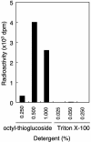
Fig. 1.
Fig. 1.
Effect of detergents on POMT activity. POMT activity was measured in a 20-μl reaction volume containing 20 mM Tris·HCl (pH 8.0), 100 nM Dol-P-[3H]Man (125,000 dpm/pmol), 2 mM 2-mercaptethanol, 10 mM EDTA, 10 μg of GST-α-DG, and 80 μg of HEK293T cell microsomal membrane fraction in the presence of n-octyl-β-d-thioglucoside or Triton X-100. The reaction was initiated by adding the protein extract and continued at 28°C for 1 h. After incubation, GST-α-DG was separated by glutathione-Sepharose beads and then the incorporated [3H]Man to GST-α-DG was measured with a liquid scintillation counter.
Fig. 2.
Fig. 2.
POMT activity in HEK293T cells. (A) Incorporation of [3H]Man into GST-α-DG. Lane 1, incubation with GST-α-DG but without membrane fraction; lane 2, incubation with membrane fraction and GST-α-DG; lane 3, incubation with membrane fraction but without GST-α-DG. (B) After incubation, a reaction mixture oflane1or2in A was subjected to SDS/PAGE (10% gel) and was stained by Coomassie. Lane 1, corresponds to lane 1 of A, and lane 2 corresponds to lane 2 of A. (C) Autoradiography of B. POMT activity was based on the rate of mannose transfer to GST-α-DG using the membrane fractions from HEK293T cells. The products were recovered by the glutathione-Sepharose beads. Incorporation of [3H]Man into GST-α-DG was measured with a liquid scintillation counter. Molecular mass standards are shown to the right of B and C.
Fig. 3.
Fig. 3.
POMT activity of human POMT1 and POMT2. Western blot analyses of myc-tagged POMT1 (A) and POMT2 (B) expressed in HEK293T cells. Lanes 1, cells transfected with vector alone; lanes 2, cells transfected with human POMT1; lanes 3, cells transfected with human POMT2; lanes 4, cells cotransfected with POMT1 and POMT2. The proteins (20 μg of HEK293T cell microsomal membrane fraction) were subjected to SDS/PAGE (10% gel), and the separated proteins were transferred to a poly(vinylidene difluoride) (PVDF) membrane. The PVDF membrane was stained with anti-myc (A) or anti-POMT2 Ab (B). Molecular mass standards are shown to the right of A and B.(C) POMT activity of membrane fractions from each of these four types (lanes 1–4) plus a mixture of the membrane fractions from the POMT1-transfected cells and POMT2-transfected cells (lane 5).
Fig. 4.
Fig. 4.
Dependence of POMT activity of human POMT1 and POMT2 on temperature (A), pH (B), and various divalent cations and EDTA (C). (A) The temperature–activity relationships at short incubation times (5–30 min) are shown (Inset). Squares, 15°C; circles, 25°C; triangles, 35°C. (B) Triangles, squares, and circles indicate sodium acetate buffer (pH 4.0∼5.5), Mes buffer (pH 5.5∼7.0), and Tris·HCl buffer (pH 7.0∼9.0), respectively. (C) The enzyme was assayed with various divalent cations at 10 mM or EDTA at 10 mM. The presence of Mg2+ had a slight enhanced effect on the activity. The enzyme was fully active in the presence of EDTA. Mn2+ and Ca2+ suppressed the enzyme activity. Eighty micrograms of HEK293T cell microsomal membrane protein was used for each assay.
Fig. 5.
Fig. 5.
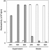
α-Mannosidase digestion of mannosyl-GST-α-DG. Glutathione-Sepharose beads bearing [3H]mannosyl-GST-α-DG were incubated with jack bean α-mannosidase. At 24-h incubation, 0.8 unit of enzyme was added at 0 h; at 48-h incubation, 0.8 unit of enzyme was added at 0 and 24 h; and at 60-h incubation, 0.8 unit of enzyme was added at 0, 24, and 48 h. After incubation and centrifugation, the radioactivities of the supernatant and the beads were measured with a liquid scintillation counter. Filled bars, active α-mannosidase; open bars, inactive (heat-treated) α-mannosidase. The radioactivity released to the supernatant increased with increasing amount of α-mannosidase used, and the radioactivity remained on the beads decreased correspondingly.

Similar articles

Cited by

References

    1. Burton, E. A. & Davies, K. E. (2002) Cell 108, 5–8. - PubMed
    1. Emery, A. E. (2002) Lancet 359, 687–695. - PubMed
    1. Muntoni, F., Brockington, M., Blake, D. J., Torelli, S. & Brown, S. C. (2002) Lancet 360, 1419–1421. - PubMed
    1. Michele, D. E. & Campbell, K. P. (2003) J. Biol. Chem. 278, 15457–15460. - PubMed
    1. Holt, K. H., Crosbie, R. H., Venzke, D. P. & Campbell, K. P. (2000) FEBS Lett. 468, 79–83. - PubMed

Publication types