. 2004 Mar;15(3):1364-73.
doi: 10.1091/mbc.e03-09-0646. Epub 2003 Dec 29.

Repression of Na,K-ATPase beta1-subunit by the transcription factor snail in carcinoma

Affiliations

Repression of Na,K-ATPase beta1-subunit by the transcription factor snail in carcinoma

Cromwell E Espineda et al. Mol Biol Cell. 2004 Mar.

Abstract

The Na,K-ATPase consists of two essential alpha- and beta-subunits and regulates the intracellular Na+ and K+ homeostasis. Although the alpha-subunit contains the catalytic activity, it is not active without functional beta-subunit. Here, we report that poorly differentiated carcinoma cell lines derived from colon, breast, kidney, and pancreas show reduced expression of the Na,K-ATPase beta1-subunit. Decreased expression of beta1-subunit in poorly differentiated carcinoma cell lines correlated with increased expression of the transcription factor Snail known to down-regulate E-cadherin. Ectopic expression of Snail in well-differentiated epithelial cell lines reduced the protein levels of E-cadherin and beta1-subunit and induced a mesenchymal phenotype. Reduction of Snail expression in a poorly differentiated carcinoma cell line by RNA interference increased the levels of Na,K-ATPase beta1-subunit. Furthermore, Snail binds to a noncanonical E-box in the Na,K-ATPase beta1-subunit promoter and suppresses its promoter activity. These results suggest that down-regulation of Na,K-ATPase beta1-subunit and E-cadherin by Snail are associated with events leading to epithelial to mesenchymal transition.


Figures
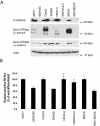
Figure 1.
Figure 1.
Correlation between E-cadherin, Na,K-ATPase α1-subunit, β1-subunit protein levels, and Na,K-ATPase enzyme activity. (A) E-cadherin, Na,K-ATPase β1-subunit, and α1-subunit protein levels were analyzed by an immunoblot in a panel of epithelial cell lines: breast, MCF7, and MDA435; colon, Caco2, and SW480; pancreas, HPAF-II and MiaPaCa-2; and canine kidney, MDCK, and MSV-transformed MDCK. Actin immunoblot analysis confirmed equal loading of whole cell lysates. MCF7, Caco2, HPAF-II, and MDCK are well-differentiated whereas MDA435, SW480, MiaPaCa-2, and MSV-MDCK are poorly differentiated cell lines. (B) Ouabain-sensitive 86Rb+ flux in well- and poorly differentiated cell lines. The 86Rb+ uptake assay was performed as described in MATERIALS AND METHODS. Bars show SE of two independent determinations performed in triplicates.
Figure 2.
Figure 2.
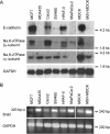
Reduced Na,K-ATPase β1-subunit mRNA levels in poorly differentiated cell lines. (A) Northern blot analysis of E-cadherin, Na,K-ATPase β1-subunit, and α1-subunit mRNA levels in well- and poorly differentiated cell lines. GAPDH Northern blot analysis confirmed equal loading of total RNA. (B) Levels of Snail mRNA expression in well- and poorly differentiated cell lines were analyzed by RT-PCR. The expression of GAPDH was analyzed in the samples as a control for the amount of cDNA present in each sample.
Figure 3.
Figure 3.
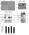
Snail expression modulates Na,K-ATPase β1-subunit levels. (A) Phase contrast image of MCF7 and MDCK cells constitutively expressing pPGS empty vector or pPGS-Snail. (B) Immunoblot analysis measured E-cadherin, Na,K-ATPase β1- and α1-subunit protein levels in MCF7 and MDCK cells constitutively expressing pPGS empty vector or pPGS-Snail. Actin immunoblot shows equal loading of whole cell lysates. (C) Ouabain-sensitive 86Rb+ flux in MCF7 and MDCK cells expressing pPGS vector or pPGS-Snail. The 86Rb+ uptake assay was performed as described in MATERIALS AND METHODS. Bars show SE of two independent determinations performed in triplicate. (D) RT-PCR analysis results show that with the exception of Na,K-ATPase α1-subunit, E-cadherin and Na,K-ATPase β1-subunit protein levels inversely correlated with Snail expression. GAPDH RT-PCR analysis was performed as a control for the amount of cDNA present in each sample. (E) RNA interference-mediated reduction of Snail increases Na,K-ATPase β1-subunit mRNA levels in SW480 cells. RNAi was performed in SW480 cells by using siRNAs for GFP (control) and Snail (refer to MATERIALS AND METHODS). After transfection, total RNA was extracted, and RT-PCR was performed using primer pairs specific for either Snail, β1-subunit, α1-subunit, E-cadherin, or GAPDH. RT-PCR products were analyzed on a 1% agarose gel.
Figure 4.
Figure 4.
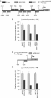
Reduced Na,K-ATPase β1-subunit promoter activity in poorly differentiated cells. (A) Schematic representation of Na,K-ATPase β1-subunit proximal promoter elements. Luciferase reporter used is under the control of the human β1-subunit promoter (Hβ1-1141-Luc). This fragment consists of half-sites of three potential MRE/GREs, four E-boxes, a nuclear factor-1 (NF-1) binding site, and a noncanonical E-box (5′ CACCGG 3′). +1 indicates the transcription start site. (B) Comparison of the β1-subunit promoter activity in well-differentiated and poorly differentiated cell lines by using the Hβ1-1141-Luc construct. MCF7, MDA435, Caco2, SW480, MDCK, and MSV-MDCK cells were transiently transfected with 10 μg of Hβ1-1141-Luc (full-length) reporter plasmid. Promoter activity was determined by luciferase reporter assays. Luciferase values were normalized to Renilla reporter activity. The results shown correspond to the average of three independent experiments. (C) Schematic representation of Hβ1-456-Luc (truncated) containing one MRE/GRE and the noncanonical E-box. (D) Comparison of the β1-subunit promoter activity in well-differentiated and poorly differentiated cell lines by using the Hβ1-456-Luc construct. MCF7, MDA435, Caco2, SW480, MDCK, and MSV-MDCK cells were transiently transfected with 10 μg of Hβ1-456-Luc reporter plasmid. Promoter activity was determined by luciferase reporter assays, and luciferase values were normalized to Renilla reporter activity. Results shown correspond to the average of three independent experiments.
Figure 5.
Figure 5.
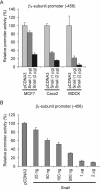
Increasing Snail expression reduces Na,K-ATPase β1-subunit promoter activity in well-differentiated cell lines (MCF7, Caco2, and MDCK) and COS cells. (A) MCF7, Caco2, and MDCK cell lines were transfected with pCDNA3 vector containing human Snail cDNA or with empty pCDNA3. One or two micrograms of pCDNA3-Snail were used in the transient transfection. The Hβ1-456-luc reporter plasmid was used in the luciferase reporter assays. Luciferase values were normalized to Renilla reporter activity. The results shown correspond to the average of three independent experiments. (B) Increasing Snail expression reduces Na,K-ATPase β1-subunit promoter activity in COS cells in a dose-dependent manner. COS cells were transfected with pCDNA3-Snail or with empty pCDNA3 plasmid. Increasing amounts of pCDNA3-Snail were used in the transient transfection. The Hβ1-456-luc plasmid was used in the luciferase reporter assays. Luciferase values were normalized to Renilla reporter activity. The results shown correspond to the average of three independent experiments.
Figure 6.
Figure 6.
Endogenous Snail from multiple cell lines binds to the noncanonical E-box element of the Na,K-ATPase β1-subunit promoter. (A) Noncanonical E-box is present at position -71 to -66 of the β1-subunit promoter. An oligonucleotide probe, called E-box1wt, was generated, which encompasses the noncanonical E-box (position -81 to -52) for the EMSA. As a control, we also generated a probe in which E-box1 is mutated into “AAATTT” (E-box1 mut). (B) Nuclear extracts from MDCK-Snail, MCF7-Snail, MiaPaCa-2, and MDA435 cells were incubated with double stranded 32P-labeled oligonucleotides containing the 5′ CACCGG 3′ sequence corresponding to E-box1 of the Na,K-ATPase β1-subunit promoter. For competition assays, a 200-fold molar excess of unlabeled E-box1 wt (wt; lanes 3, 7, 12, and 16), E-box1 mut (mut; lanes 4, 8, 13, and 17) oligonucleotides, 8 μg of anti-SNAI-1 antibody (Snail; lanes 5, 9, 14, and 18) or a nonspecific antibody (Cont; lane 10) was added before addition of probe to the binding reactions. Snail-containing complexes were detected in all cell lines (arrow). Addition of anti-SNAI-1 antibody or cold wild-type probe competed Snail binding to E-box1.

Similar articles

Cited by

References

    1. Abbott, A., and Ball, W.J., Jr. (1993). The epitope for the inhibitory antibody M7-PB-E9 contains Ser-646 and Asp-652 of the sheep Na+, K(+)-ATPase alpha-subunit. Biochemistry 32, 3511-3518. - PubMed
    1. Batlle, E., Sancho, E., Franci, C., Dominguez, D., Monfar, M., Baulida, J., and Garcia De Herreros, A. (2000). The transcription factor snail is a repressor of E-cadherin gene expression in epithelial tumour cells. Nat. Cell Biol. 2, 84-89. - PubMed
    1. Beguin, P., Wang, X., Firsov, D., Puoti, A., Claeys, D., Horisberger, J.D., and Geering, K. (1997). The gamma subunit is a specific component of the Na, K-ATPase and modulates its transport function. EMBO J. 16, 4250-4260. - PMC - PubMed
    1. Behrens, J., Mareel, M.M., Van Roy, F.M., and Birchmeier, W. (1989). Dissecting tumor cell invasion: epithelial cells acquire invasive properties after the loss of uvomorulin-mediated cell-cell adhesion. J. Cell Biol. 108, 2435-2447. - PMC - PubMed
    1. Blanco, G., Melton, R.J., Sanchez, G., and Mercer, R.W. (1999). Functional characterization of a testes-specific alpha-subunit isoform of the sodium/potassium adenosinetriphosphatase. Biochemistry 38, 13661-13669. - PubMed

Publication types

MeSH terms