. 2004 Mar;15(3):1146-59.
doi: 10.1091/mbc.e03-08-0581. Epub 2003 Dec 29.

Depletion of centromeric MCAK leads to chromosome congression and segregation defects due to improper kinetochore attachments

Affiliations

Depletion of centromeric MCAK leads to chromosome congression and segregation defects due to improper kinetochore attachments

Susan L Kline-Smith et al. Mol Biol Cell. 2004 Mar.

Abstract

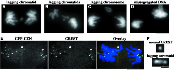
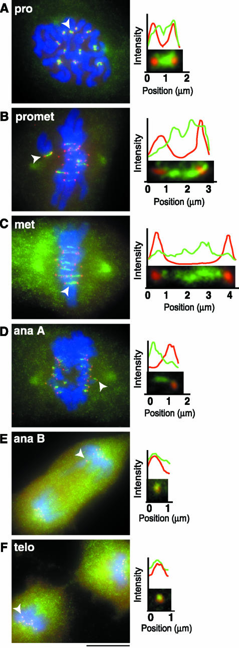
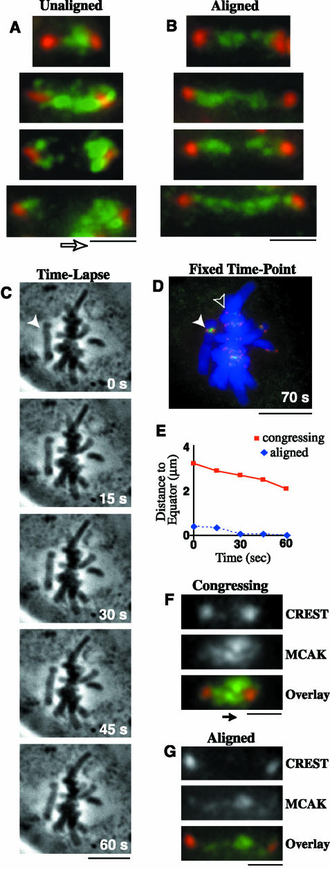
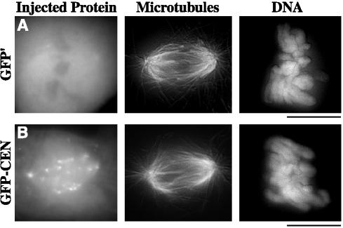
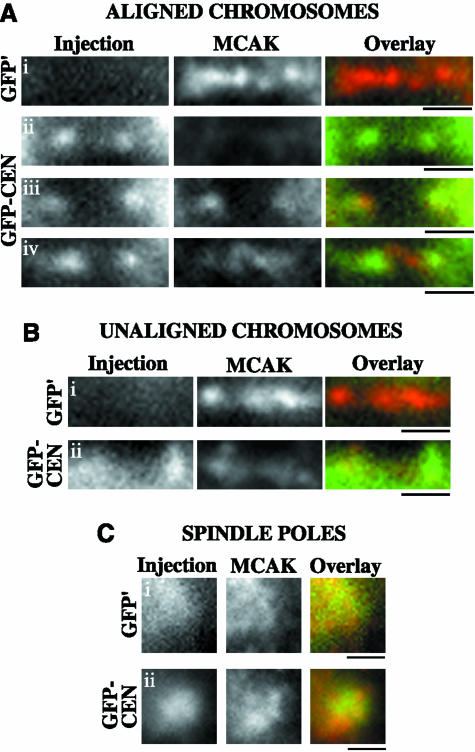
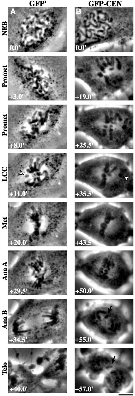
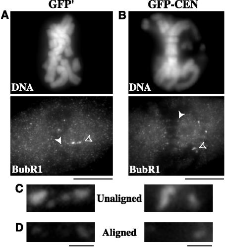
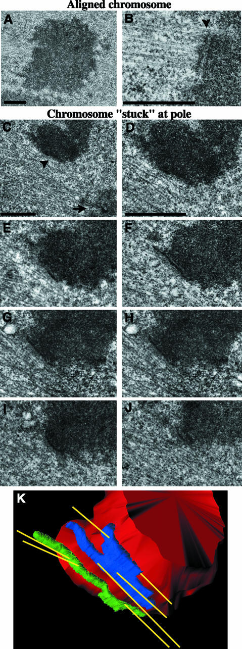
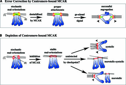
The complex behavior of chromosomes during mitosis is accomplished by precise binding and highly regulated polymerization dynamics of kinetochore microtubules. Previous studies have implicated Kin Is, unique kinesins that depolymerize microtubules, in regulating chromosome positioning. We have characterized the immunofluorescence localization of centromere-bound MCAK and found that MCAK localized to inner kinetochores during prophase but was predominantly centromeric by metaphase. Interestingly, MCAK accumulated at leading kinetochores during congression but not during segregation. We tested the consequences of MCAK disruption by injecting a centromere dominant-negative protein into prophase cells. Depletion of centromeric MCAK led to reduced centromere stretch, delayed chromosome congression, alignment defects, and severe missegregation of chromosomes. Rates of chromosome movement were unchanged, suggesting that the primary role of MCAK is not to move chromosomes. Furthermore, we found that disruption of MCAK leads to multiple kinetochore-microtubule attachment defects, including merotelic, syntelic, and combined merotelic-syntelic attachments. These findings reveal an essential role for Kin Is in prevention and/or correction of improper kinetochore-microtubule attachments.


Figures

Figure 7.
Figure 7.
Depletion of centromere-bound MCAK leads to severe chromosome missegregation. PtK2 cells in prophase were injected with GFP-CEN and processed for fluorescence microscopy of DNA at 40 min postinjection. Segregation defects in GFP-CEN–injected cells included (A and B) lagging chromatids during anaphase and telophase, (C) lagging chromosomes during anaphase, as well as (B and D) grossly missegregated amounts of DNA. (E and F) GFP-CEN–injected cells were fixed and immunostained for CREST and DNA at 40 min postinjection. (E) GFP-CEN–injected cell with lagging chromatid (white arrow) and missegregated DNA in anaphase B. CREST staining shows that 17 chromatids were segregated to the left pole, whereas 11 chromatids were segregated to the right pole. (F) The CREST staining pattern of the lagging chromosome (bottom) is stretched threefold compared with CREST at a normally segregated chromatid (top), indicated by arrowhead in E. Bars (A-E), 10 μm; (F), 1 μm.
Figure 1.
Figure 1.
MCAK displays differential localization to centromeres throughout mitosis. (A–F, left column) PtK2 cells were fixed and stained for CREST (red), MCAK (green), and DNA (blue). To analyze the extent of colocalization of MCAK and CREST, line scans were generated in MetaMorph by drawing a two-pixel width line through centromeres for each stage of mitosis (A–F, right column). Centromeres used to generate line scans in this figure are denoted by solid arrowheads. Bar, 10 μm.
Figure 2.
Figure 2.
MCAK is enriched at the leading kinetochore of congressing chromosomes. PtK2 cells were fixed and immunostained for CREST (red), MCAK (green), and DNA (blue). (A) Centromeres of unaligned chromosomes exhibited increased levels of MCAK at the kinetochore facing the metaphase plate (direction indicated by open arrow), whereas (B) centromeres of aligned chromosomes from the same cells showed an equal distribution of MCAK at both sister kinetochores. (C) Congressing chromosomes were analyzed by time-lapse phase contrast microscopy at 15-s intervals. Actual times are given in the lower right corner. White arrowhead indicates a chromosome moving to the metaphase plate for 1 min. (D) The cell shown in part C was fixed and immunostained to analyze a congressing chromosome (solid arrowhead), which exhibited motion toward the metaphase plate at 1.1 μm/min (E; solid red line), and displayed an increase in MCAK staining at the leading kinetochore (F; direction of motion indicated by the solid arrow). The aligned chromosome in D (open arrowhead), exhibited negligible motion at the metaphase plate (E; dashed blue line), and showed no difference in MCAK staining at either sister kinetochore (G). Bars (A, B, F, and G), 1 μm; (C and D), 10 μm.
Figure 3.
Figure 3.
GFP-CEN binds kinetochores, centromeres, and spindle poles without disrupting global spindle morphology. PtK2 cells in prophase were injected with GFP′ control (A) or GFP-CEN (B), the centromere dominant-negative version of MCAK. At 30 min postinjection, cells were fixed and immunostained for MTs and DNA. Bars, 10 μm.
Figure 4.
Figure 4.
GFP-CEN depletes endogenous MCAK from centromeres but not from spindle poles. PtK2 cells in prophase were injected with GFP′ control or GFP-CEN protein. At 30′ postinjection, cells were fixed and stained to visualize endogenous MCAK (red) versus the injected proteins (green). Cells were analyzed by Z-series optical sectioning, and single plane images are shown. (A, i) Aligned chromosomes in GFP′-injected cells exhibited normal endogenous MCAK localization to the centromere. GFP-CEN binding at kinetochores inhibited endogenous MCAK from localizing to centromeres in the majority of cells (A, ii and iii), although a few aligned chromosomes retained a small amount of MCAK at the centromere (A, iv). Unaligned chromosomes in GFP′-injected cells exhibited intense localization of endogenous MCAK at the centromere (B, i), whereas GFP-CEN binding decreased the localization of MCAK (B, ii). Although both GFP′ and GFP-CEN bound spindle poles during mitosis (C, i and ii), neither fusion protein disrupted MCAK localization to the spindle pole. Bars, 1 μm.
Figure 5.
Figure 5.
Depletion of centromere-bound MCAK perturbs chromosome alignment and segregation. PtK2 cells in prophase were injected with (A) GFP′ or (B) GFP-CEN. Time-lapse phase contrast microscopy was used to image cells as they progressed through mitosis. The time for each stage of mitosis is given in minutes (bottom left), with NEB at t = 0.0 min. LCC is the time at which the last chromosome began congression (arrowheads). Note lagging chromatid (arrows) during anaphase B and telophase in B. Bar, 10 μm.
Figure 6.
Figure 6.
BubR1 stains kinetochores of unaligned chromosomes but not aligned chromosomes in GFP-CEN–injected PtK2 cells. BubR1 staining at kinetochores was compared between GFP′-injected control cells (A) and GFP-CEN–injected cells (B). (C) BubR1 staining at kinetochores of unaligned chromosomes is indicated in A and B by open arrowheads. (D) BubR1 staining at kinetochores of aligned chromosomes is indicated in A and B by solid arrowheads. Bars (A and B), 10 μm; (C and D), 1 μm.
Figure 8.
Figure 8.
Centromeric MCAK depletion leads to improper kinetochore–microtubule attachments in PtK2 cells. Correlative light/serial-section EM was performed on GFP-CEN–injected cells to analyze kinetochore and K-fiber morphology. Low-magnification (A) and high-magnification (B) images of an aligned chromosome with normal kinetochore morphology and MT attachments; arrowhead is positioned at the kinetochore–MT interface. (C) Low-magnification image of a chromosome that remained for an extended period of time at the pole during mitosis. Arrow denotes a centrosome; arrowhead denotes a kinetochore. (D–J) High-magnification, 100-nm serial-section images of the chromosome shown in C reveal that both kinetochores are attached to MTs emanating from both poles, which makes this chromosome both merotelically and syntelically oriented on the spindle. (K) The 3-D structure of MTs attached to the merotelic-syntelic kinetochores of the chromosome shown in C–J. The chromosome is shown in red, MTs in yellow, and sister kinetochores in blue and green. Bars, 1 μm.
Figure 9.
Figure 9.
Model for destabilization of maloriented kinetochore–microtubule attachments by centromere-bound MCAK. Chromosomes are in blue, CREST is in red, MCAK is in green, and MCAK colocalized with CREST is in yellow. (A) Maloriented MT attachments, such as kinetochore MTs penetrating past the outer kinetochore (1) or nonkinetochore MTs invading the centromere (2) are depolymerized upon encountering MCAK. This allows for turnover and the eventual formation of properly oriented attachments. The spindle checkpoint gives the go-ahead signal, and proper segregation is achieved. (B) On depletion of centromere-bound MCAK, however, maloriented attachments are not destabilized. Stable malorientations (such as merotelic and/or syntelic kinetochore–MT attachments) are not resolved, and missegregation of chromatids occurs at anaphase.

Similar articles

Cited by

References

    1. Biggins, S., Severin, F.F., Bhalla, N., Sassoon, I., Hyman, A.A., and Murray, A.W. (1999). The conserved protein kinase Ipl1 regulates microtubule binding to kinetochores in budding yeast. Genes Dev. 13, 532-544. - PMC - PubMed
    1. Cassimeris, L., Rieder, C.L., Rupp, G., and Salmon, E.D. (1990). Stability of microtubule attachment to metaphase kinetochores in PtK1 cells. J. Cell Sci. 96, 9-15. - PubMed
    1. Cassimeris, L., Rieder, C.L., and Salmon, E.D. (1994). Microtubule assembly and kinetochore directional instability in vertebrate monopolar spindles: implications for the mechanism of chromosome congression. J. Cell Sci. 107, 285-297. - PubMed
    1. Cimini, D., Howell, B., Maddox, P., Khodjakov, A., Degrassi, F., and Salmon, E.D. (2001). Merotelic kinetochore orientation is a major mechanism of aneuploidy in mitotic mammalian tissue cells. J. Cell Biol. 153, 517-527. - PMC - PubMed
    1. Cimini, D., Fioravanti, D., Salmon, E.D., and Degrassi, F. (2002). Merotelic kinetochore orientation versus chromosome mono-orientation in the origin of lagging chromosomes in human primary cells. J. Cell Sci. 115, 507-515. - PubMed

Publication types