. 2004 Mar;15(3):1273-86.
doi: 10.1091/mbc.e03-07-0491. Epub 2003 Dec 29.

Tumor necrosis factor-alpha promotes survival of opossum kidney cells via Cdc42-induced phospholipase C-gamma1 activation and actin filament redistribution

Affiliations

Tumor necrosis factor-alpha promotes survival of opossum kidney cells via Cdc42-induced phospholipase C-gamma1 activation and actin filament redistribution

Evangelia A Papakonstanti et al. Mol Biol Cell. 2004 Mar.

Abstract

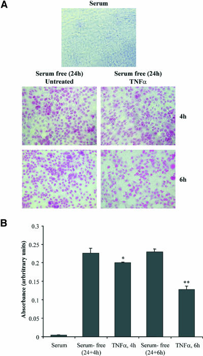
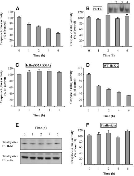
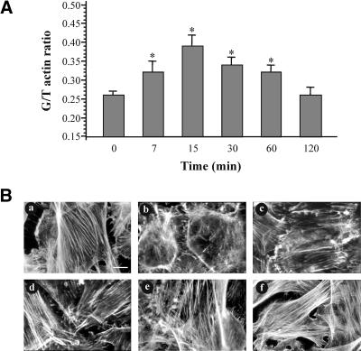
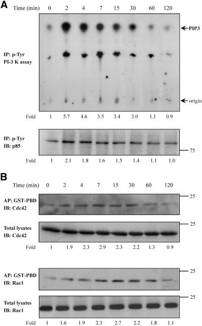
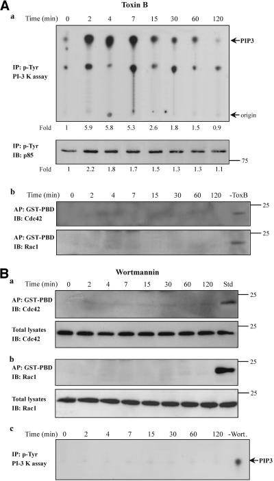
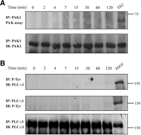
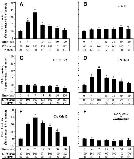
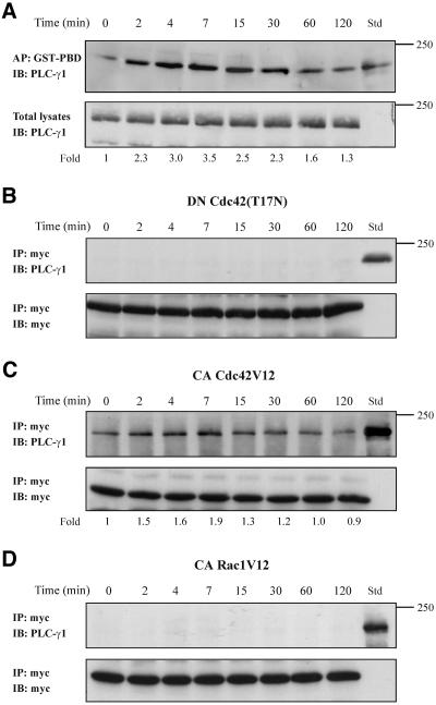
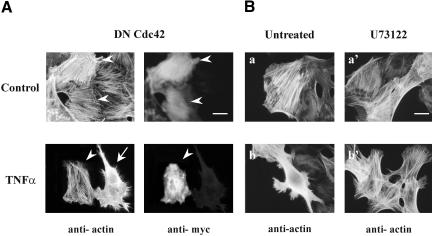
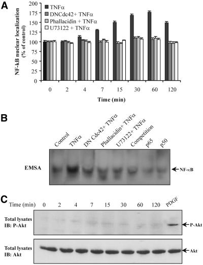
Although the renal proximal tubular epithelial cells are targeted in a variety of inflammatory diseases of the kidney, the signaling mechanism by which tumor necrosis factor (TNF)-alpha exerts its effects in these cells remains unclear. Here, we report that TNF-alpha elicits antiapoptotic effects in opossum kidney cells and that this response is mediated via actin redistribution through a novel signaling mechanism. More specifically, we show that TNF-alpha prevents apoptosis by inhibiting the activity of caspase-3 and this effect depends on actin polymerization state and nuclear factor-kappaB activity. We also demonstrate that the signaling cascade triggered by TNF-alpha is governed by the phosphatidylinositol-3 kinase, Cdc42/Rac1, and phospholipase (PLC)-gamma1. In this signaling cascade, Cdc42 was found to be selectively essential for PLC-gamma1 activation, whereas phosphatidylinositol-3,4,5-triphosphate alone is not sufficient to activate the phospholipase. Moreover, PLC-gamma1 was found to associate in vivo with the small GTPase(s). Interestingly, PLC-gamma1 was observed to associate with constitutively active (CA) Cdc42V12, but not with CA Rac1V12, whereas no interaction was detected with Cdc42(T17N). The inactive Cdc42(T17N) and the PLC-gamma1 inhibitor U73122 prevented actin redistribution and depolymerization, confirming that both signaling molecules are responsible for the reorganization of actin. Additionally, the actin filament stabilizer phallacidin potently blocked the nuclear translocation of nuclear factor-kappaB and its binding activity, resulting in abrogation of the TNF-alpha-induced inhibition of caspase-3. To conclude, our findings suggest that actin may play a pivotal role in the response of opossum kidney cells to TNF-alpha and implicate Cdc42 in directly regulating PLC-gamma1 activity.


Figures

Figure 1.
Figure 1.
Antiapoptotic effects of TNF-α in OK cells. Cells in complete medium or cells that had been exposed to serum-free medium for 24 h and then incubated with TNF-α for the indicated times were assessed for apoptosis using the colorimetric APOPercentage apoptosis assay. Apoptotic cells were photographed using an inverted microscope (A) or quantified by measuring the absorbance by using a microplate colorimeter (B). Mean + SEM from two separate experiments performed in triplicate (significance level *p < 0.05, **p < 0.01).
Figure 2.
Figure 2.
Involvement of NF-κB and actin in the inhibition of caspase-3 by TNF-α. Extracts of TNF-α-treated cells (A) or cells that had been preincubated with PDTC (B), or had been transfected with IκBα(S32A/S36A) (C) or WT IKK-2 (D), or had been preincubated with phallacidin (F) and then treated with TNF-α, for the indicated times, were incubated with the caspase-3 substrate DEVD-p-nitroanilide, and the protease activity was analyzed as described in MATERIALS AND METHODS. Data are presented as percentage of control cells' activity by measuring the OD per milligram of protein per minute (mean + SEM from two to three separate experiments performed in duplicate). (B, inset) Cells were incubated with TNF-α for 1 h in the presence or absence of PDTC. Nuclear extracts were subjected to EMSA as described in MATERIALS AND METHODS. Lane 1, untreated cells; lane 2, cells treated with PDTC; lane 3, cells treated with TNF-α; lane 4, cells treated with TNF-α in the presence of PDTC. (E) Cells were stimulated with TNF-α for the indicated times. Total cell lysates were prepared and immunoblotted using anti-Bcl-2 (top) or anti-actin antibody (bottom).
Figure 3.
Figure 3.
Actin redistribution and depolymerization in response to TNF-α. (A) Cells were incubated for the indicated times with 10 ng/ml TNF-α and then the monomeric (G) or total (T) actin levels was measured as described in MATERIALS AND METHODS. Data are presented as G/T actin ratio (mean + SEM from five separate experiments; significance level *p < 0.05). (B) Cells were incubated with TNF-α for 7 (b), 15 (c), 30 (d), 60 (e), or 120 (f) min and then the redistribution of filamentous actin was determined with rhodamine-phalloidin staining by immunofluorescence microscopy. (a) Untreated cells. Bar, 20 μm. Similar results were obtained in three independent experiments.
Figure 4.
Figure 4.
Effect of TNF-α on the PI-3 kinase and Cdc42/Rac1 activity. Cells were incubated for the indicated times with TNF-α (10 ng/ml). Equal amount of proteins of cell lysates were immunoprecipitated with an anti-phosphotyrosine antibody and subjected to an in vitro PI-3 kinase assay, as described in MATERIALS AND METHODS, by using PIP2 as substrate. The number below each lane indicates the fold amount of phosphatidylinositol-3,4,5-trisphosphate (PIP3) product, with that of untreated cells taken as 1 (top). The phosphorylation of the p85 regulatory subunit of PI-3 kinase that was immunoprecipitated in the kinase assay was assessed by immunoprecipitation (IP) with an anti-phosphotyrosine antibody and immunoblotting (IB) with anti-PI-3 kinase (p85) antibody. The number below each lane indicates the fold phosphorylation of p85, with that of untreated cells taken as 1 (bottom). (B) Equal volume of cell lysates from untreated or TNF-α-treated cells were affinity precipitated (AP) with GTP-PBD bound to glutathione-agarose beads. Precipitated GTP-Cdc42 or GTP-Rac1 was detected by IB with anti-Cdc42 or anti-Rac1 antibody, respectively. Equal volume of total lysates from untreated and TNF-α-treated cells were subjected to SDS-PAGE, transferred to nitrocellulose membrane, and IB with monoclonal anti-Cdc42 or anti-Rac1 antibody, respectively. The number below each lane indicates the normalized fold activation of Cdc42 or Rac1, with that of untreated cells taken as 1. Results shown are representative of three independent experiments with similar results.
Figure 5.
Figure 5.
PI-3 kinase is activated upstream of the small GTPases Cdc42/Rac1 in TNF-α-treated cells. (A) Cells were pretreated with toxin B (50 ng/ml, 2 h) and then incubated with TNF-α (10 ng/ml) for the indicated times. (a) Equal amount of proteins of cell lysates were immunoprecipitated with an anti-phosphotyrosine antibody and subjected to an in vitro PI-3 kinase assay, as described in MATERIALS AND METHODS, by using PIP2 as substrate. The number below each lane indicates the fold amount of PIP3 product, with that of untreated cells taken as 1 (top). The phosphorylation of the p85 regulatory subunit of PI-3 kinase that was immunoprecipitated in the kinase assay was assessed by immunoprecipitation (IP) with an anti-phosphotyrosine antibody and immunoblotting (IB) with anti-PI-3 kinase (p85) antibody. The number below each lane indicates the fold phosphorylation of p85, with that of untreated cells taken as 1 (bottom). (b) The activated forms of Cdc42 (top) or Rac1 (bottom) in the presence or absence (-ToxB) of toxin B was determined by affinity precipitation (AP) with GST-PBD and then by IB with the respective antibodies. (B) Cells were pretreated with wortmannin (100 nM, 30 min) and then with TNF-α. Equal volume of cell lysates from untreated or TNF-α-treated cells were affinity precipitated (AP) with GTP-PBD bound to glutathione-agarose beads. Precipitated GTP-Cdc42 (a, top) or GTP-Rac1 (b, top) was detected by immunoblot (IB) with anti-Cdc42 or anti-Rac1 antibody, respectively. Equal volumes of total lysates from untreated and TNF-α-treated cells were subjected to SDS-PAGE, transferred to nitrocellulose membrane, and IB with monoclonal anti-Cdc42 (a, bottom) or anti-Rac1 (a, bottom) antibody, respectively. (c) The lipid kinase activity of PI-3 kinase in the presence or absence (-Wort.) of wortmannin was determined by the in vitro PI-3 kinase assay, as described in MATERIALS AND METHODS, by using PIP2 as substrate. Results shown are representative of three similar experiments. PIP3, phosphatidylinositol-3,4,5-trisphosphate.
Figure 6.
Figure 6.
TNF-α has no effect on PAK1 activity and on PLC-γ1 phosphorylation. (A) Cells were stimulated with TNF-α for the indicated times or with 10-8 M EKC for 30 min (positive control), lysed, and anti-PAK1 immune complexes were assayed for kinase autophosphorylation by an in vitro kinase assay as described in MATERIALS AND METHODS. The reaction products were separated by SDS-PAGE, transferred to nitrocellulose membrane, and phosphorylation was visualized by autoradiography (top). The amount of PAK1 protein that was immunoprecipitated in the kinase assay was assessed by immunoblotting (IB) with anti-PAK1 antibody (bottom). (B) Cells were stimulated with TNF-α for the indicated times or with 40 ng/ml platelet-derived growth factor for 15 min (positive control), lysed, and then equal amount of proteins were immunoprecipitated (IP) with an anti-phosphotyrosine antibody. The tyrosine-phosphorylated PLC-γ1 was detected by IB with a specific anti-PLC-γ1 antibody (top). PLC-γ1 phosphorylation was also examined by its immunoprecipitation that was followed by immunoblotting with an anti-phosphotyrosine antibody (middle). Stripping and reprobing of the nitrocellulose membrane confirmed the presence of PLC-γ1 protein on immunoprecipitates (bottom). Results shown are representative of three similar experiments.
Figure 7.
Figure 7.
PLC-γ1 activity depends on Cdc42 activation in TNF-α-treated cells. PLC-γ1 was immunoprecipitated from equal amount of proteins from TNF-α-treated cells (A) or cells that had been preincubated with toxin B (B) or had been transfected with Cdc42(T17N) (C), Rac1(T17N) (D), Cdc42V12 (E), or with Cdc42V12 followed by exposure to wortmannin (100 nM, 30 min) (F) and then incubated with TNF-α for the indicated times. PLC-γ1 was analyzed for hydrolytic activity toward [3H]phosphatidylinositol 4,5-bisphosphate as described in MATERIALS AND METHODS. PLC-γ1 activity is presented as percentage of control cells' activity by measuring the cpm [3H]inositol 1,4,5-triphosphate released (mean + SEM from three separate experiments performed in duplicate, *p < 0.05). To confirm that equal amounts of PLC-γ1 protein were assayed under each condition separate immunoblot experiments were performed. The “OD × area” (as percentage of control) (±SEM) corresponding to bands of PLC-γ1 loaded was measured by PC-based image analysis and presented in table below each panel.
Figure 8.
Figure 8.
PLC-γ1 associates in vivo with Cdc42. (A) Equal volumes of cell lysates from untreated or TNF-α-treated cells were affinity precipitated (AP) with GTP-PBD bound to glutathione-agarose beads. Coprecipitated PLC-γ1 was detected by immunoblot (IB) with anti-PLC-γ1 antibody. Total lysates were used as Std to assess the mobility of precipitated PLC-γ1 (top). Equal volume of total lysates from untreated and TNF-α-treated cells were subjected to SDS-PAGE, transferred to nitrocellulose membrane, and immunoblotted (IB) with anti-PLC-γ1 antibody (bottom). The number below each lane indicates the normalized fold amount of PLC-γ1 precipitated with GST-PBD, with that of untreated cells taken as 1. (B) Cells were transfected with the inactive Cdc42(T17N) and then stimulated with TNF-α for the indicated times. Myc-tagged Cdc42(T17N) was immunoprecipitated (IP) with mouse monoclonal anti-myc epitope antibody, and Western blot of immunoprecipitates was probed (IB) with anti-PLC-γ1 antibody. Total lysates were used as Std to assess the mobility of precipitated PLC-γ1 (top). The blot was stripped and reprobed with anti-myc antibody to confirm the presence of Cdc42(T17N) in the immunoprecipitates (bottom). (C) Cells were transfected with the constitutively active Cdc42V12 and then stimulated with TNF-α for the indicated times. Myc-tagged Cdc42V12 was IP with mouse monoclonal anti-myc epitope antibody and western blot of immunoprecipitates was probed (IB) with anti-PLC-γ1 antibody. Total lysates were used as Std to assess the mobility of precipitated PLC-γ1 (top). The blot was stripped and reprobed with anti-myc antibody to confirm the presence of Cdc42V12 in the immunoprecipitates (bottom). The number below each lane indicates the fold amount of PLC-γ1 coimmunoprecipitated with Cdc42V12, with that of untreated cells taken as 1. (D) Cells were transfected with the constitutively active Rac1V12 and then stimulated with TNF-α for the indicated times. Myc-tagged Rac1V12 was IP with mouse monoclonal anti-myc epitope antibody and Western blot of immunoprecipitates was probed (IB) with anti-PLC-γ1 antibody. Total lysates were used as Std to assess the mobility of precipitated PLC-γ1 (top). The blot was stripped and reprobed with anti-myc antibody to confirm the presence of Rac1V12 in the immunoprecipitates (bottom). The experiments were repeated four times with similar results.
Figure 9.
Figure 9.
The TNF-α-induced actin remodeling occurs through a Cdc42- and PLC-γ1-dependent mechanism. Cells were transfected with Myc-tagged dominant negative Cdc42(T17N) (A) or preincubated with U73122 (B) and then incubated with TNF-α for 15 min. Transfected cells were identified by double immunofluorescence as described in MATERIALS AND METHODS. Cells expressing Myc-tagged Cdc42(T17N) are indicated by an arrowhead; arrows show untransfected cells. Bar, 30 μm. Similar results were obtained in three independent experiments.
Figure 10.
Figure 10.
NF-κB nuclear translocation is dependent on actin polymerization state. (A) Nuclear extracts of cells incubated with TNF-α or cells that had been transfected with Cdc42(T17N) or that had been preincubated with phallacidin or U73122 and then exposed to TNF-α, for the indicated times, were prepared as described in MATERIALS AND METHODS. The nuclear levels of NF-κB (p65) protein were immunodetected using a specific anti-NF-κB (p65) antibody, and the band's intensity was quantified using a PC-based image analysis. Data are presented as percentage of NF-κB (p65) protein levels in control cells (mean + SEM from three separate experiments). (B) Cells were incubated with TNF-α for 30 min. Nuclear extracts were subjected to EMSA as described in MATERIALS AND METHODS. Specificity of the bands was determined using anti-p65 (p65) or anti-p50 (p50) antibodies or by competition with 25× excess of unlabeled probe. (C) Cells were treated with TNF-α, for the indicated times, or with platelet-derived growth factor (PDGF) (40 ng/ml, 15 min) that was used as positive control and then equal amount of proteins (50 μg) were subjected to SDS-PAGE, transferred to nitrocellulose membrane, and immunoblotted (IB) with an anti-phospho-Akt (top) or anti-Akt (bottom) antibody.

Similar articles

Cited by

References

    1. Are, A.F., V. E., Galkin, T. V. Pospelova, and G. P. Pinaev. (2000). The p65/RelA subunit of NF-kappaB interacts with actin-containing structures. Exp. Cell Res. 256, 533-544. - PubMed
    1. Arkinstall, S., Payton, M., and Maundrell, K. (1995). Activation of phospholipase C gamma in Schizosaccharomyces pombe by coexpression of receptor or nonreceptor tyrosine kinases. Mol. Cell. Biol. 15, 1431-1438. - PMC - PubMed
    1. Auger, K.R., Serunian, L.A., Soltoff, S.P., Libby, P., and Cantley, C. (1989). PDGF-dependent tyrosine phosphorylation stimulates production of novel polyphosphoinositides in intact cells. Cell 57, 167-175. - PubMed
    1. Bae, Y.S., Cantley, L.G., Chen, C.S., Kim, S.R., Kwon, K.S., and Rhe, S.G. (1998). Activation of phospholipase C-gamma by phosphatidylinositol 3, 4, 5-trisphosphate. J. Biol. Chem. 273, 4465-4469. - PubMed
    1. Bagrodia, S., Taylor, S.J., Creasy, C.L., Chernoff, J., and Cerione, R.A. (1995). Identification of a mouse p21Cdc42/Rac-activated kinase. J. Biol. Chem. 270, 22731-22737. - PubMed

Publication types

MeSH terms