. 2004 Mar;15(3):982-9.
doi: 10.1091/mbc.e03-06-0359. Epub 2003 Dec 29.

Nonmuscle myosin IIb is involved in the guidance of fibroblast migration

Collaborators, Affiliations

Nonmuscle myosin IIb is involved in the guidance of fibroblast migration

Chun-Min Lo et al. Mol Biol Cell. 2004 Mar.

Abstract

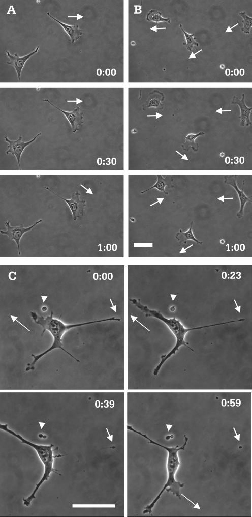
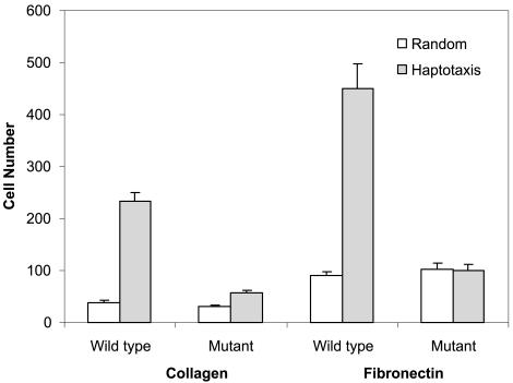
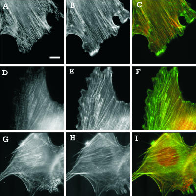
Although myosin II is known to play an important role in cell migration, little is known about its specific functions. We have addressed the function of one of the isoforms of myosin II, myosin IIB, by analyzing the movement and mechanical characteristics of fibroblasts where this protein has been ablated by gene disruption. Myosin IIB null cells displayed multiple unstable and disorganized protrusions, although they were still able to generate a large fraction of traction forces when cultured on flexible polyacrylamide substrates. However, the traction forces were highly disorganized relative to the direction of cell migration. Analysis of cell migration patterns indicated an increase in speed and decrease in persistence, which were likely responsible for the defects in directional movements as demonstrated with Boyden chambers. In addition, unlike control cells, mutant cells failed to respond to mechanical signals such as compressing forces and changes in substrate rigidity. Immunofluorescence staining indicated that myosin IIB was localized preferentially along stress fibers in the interior region of the cell. Our results suggest that myosin IIB is involved not in propelling but in directing the cell movement, by coordinating protrusive activities and stabilizing the cell polarity.


Figures

Figure 1.
Figure 1.
Morphology and migratory behavior of control and myosin IIB null cells. Although most control cells maintained their morphology over a 60-min period (A), mutant cells showed rapid changes in cell shape during the same period of time (B). In addition mutant cells migrate with a highly unstable polarity (B, arrows), compared with control cells (A, arrows). Images of high magnification show protrusion, elongation (C, long arrows), and rapid retraction of processes (C, short arrows). The latter creates small pieces of motile cytoplasm that litter the glass surface (C, arrowheads). Time in hours and minutes is indicated in each image. Bar, 50 μm.
Figure 2.
Figure 2.
Restoration of normal morphology of myosin IIB mutant cells by the reexpression of GFP-myosin IIB. Myosin IIB nulls were transfected with a vector carrying the sequence of GFP-myosin IIB. GFP signals were found along stress fibers (A). Cells expressing GFP-myosin IIB show a normal morphology and no random protrusive activities (B and C). In contrast, cells expressing GFP-myosin IIA maintained the irregular, unstable morphology as for the original myosin IIB null cells (D and E). Time in hours and minutes is indicated. Bar, 50 μm.
Figure 3.
Figure 3.
Quantitative analysis of cell migration. Plotting of mean squared distance against time (from 11 control cells and 17 mutant cells; each data point represents a mean ± SEM) indicates that mutant cells (A, open square) are able to migrate over a longer distance than do control cells (A, solid square), irrespective of the direction. However, double reciprocal plot of root mean squared distance against time, where migration speed is calculated as 1/slope and directional persistence is calculated as slope/6× y-intercept, indicates that mutant cells (B, open square) migrate at a higher speed but with a decreased persistence compared with control cells (B, solid square; Table 1).
Figure 4.
Figure 4.
Haptotactic migration assay of control and myosin IIB mutant cells. To measure haptotactic movements, cells were plated on porous membrane with one side coated with collagen or fibronectin, at a concentration of 40 and 20 μg/ml, respectively. Random migration was measured by applying this coating to both sides of the membrane. The number of cells that migrated across the membrane was counted after 15 h. Bars represent the mean ± SE from six independent experiments. For either collagen or fibronectin coating, myosin IIB null cells show no significant haptotactic migration. To the contrary, control cells show a haptotaxis/random migration ratio of 5–6.
Figure 5.
Figure 5.
Wound-induced migration of control and myosin IIB null cells. Confluent monolayers of either control (A) or myosin IIB null (B) cells were wounded at time 0. Control cells migrate and cover the cell-free area in ∼6 h, whereas mutant cells cover only about two-thirds of the area over the same period. Hours and minutes since the wound are indicated in each image. Bar, 100 μm. Average rates of wound closure during the first 4 h of repair were calculated from six independent experiments (C). Error bars indicate SE of the mean.
Figure 6.
Figure 6.
Traction forces generated by control (A and B) and myosin IIB null cells (C and D). Cells were plated on flexible substrates embedded with beads. The distribution of traction stress was calculated based on the displacements of beads and rendered as vector plots (A and C). Graphs show the corresponding angular distribution of traction forces at four time points (B and D). Control cells show a pattern of traction stress similar to that of 3T3 fibroblasts (A), with strong traction forces concentrated at the leading edge. The direction of maximal traction force (A, short hollow arrow) is antiparallel to the direction of cell migration (A, long hollow arrow) and is 90° from the direction of minimal traction forces (B). In contrast, mutant cells show an unstable, poorly defined direction of maximal traction forces (C, short hollow arrow; D), which bears no apparent relationship to either the direction of cell migration (C, long hollow arrow) or the direction of minimal traction force (D).
Figure 7.
Figure 7.
Response of myosin IIB null cells to local mechanical stimulations. A blunted microneedle was inserted into the polyacrylamide substrate near the lateral edge of a cell and then moved toward the cell to compress the substrate (arrows). The local change in substrate tension did not cause the mutant cell to change its direction of movement (B, arrowheads). However, similar manipulations caused control cells to move away from the needle (A, arrowheads). Hours and minutes since the beginning of the manipulation are indicated in each image. Bar, 50 μm.
Figure 8.
Figure 8.
Organization of myosin IIA and IIB in control and myosin IIB null cells. Control cells (A–F) or myosin IIB null cells (G–I) were stained with antibodies against myosin IIA (A and G) or myosin IIB (D) and counterstained with fluorescent phalloidin (B, E, and H). Merged images of myosin and actin (C, F, and I) show regions enriched (more red) or depleted (more green) of the specific isoform of myosin. Myosin IIA seems to be more evenly distributed among stress fibers in central and peripheral regions (A–C and G–I), whereas myosin IIB is localized preferentially along stress fibers in the central region of control cells (D–F). Moreover, myosin IIA shows a more discrete punctate pattern along stress fibers than does myosin IIB (A and G). Bar, 10 μm.

References

    1. Anderson, K.I., Wang, Y.-L., and Small, J.V. (1996). Coordination of protrusion and translocation of the keratocyte involves rolling of the cell body. J. Cell Biol. 134, 1209-1218. - PMC - PubMed
    1. Berg, J.S., Powell, B.C., and Cheney, R.E. (2001). A millennial myosin census. Mol. Biol. Cell 12, 780-94. - PMC - PubMed
    1. Bridgman, P.C., Dave, S., Asnes, C.F., Tullio, A.N., and Adelstein, R.S. (2001). Myosin IIB is required for growth cone motility. J. Neurosci. 21, 6159-6169. - PMC - PubMed
    1. Brown, M.E., and Bridgman, P.C. (2002). Retrograde flow rate is increased in growth cones from myosin IIB knockout mice. J. Cell Sci. 116, 1087-1094. - PubMed
    1. Burridge, K., and Chrzanowska-Wodnicka, M. (1996). Focal adhesions, contractility, and signaling. Annu. Rev. Cell. Dev. Biol. 12, 463-518. - PubMed

Publication types