. 2004 Jan 5;164(1):19-24.
doi: 10.1083/jcb.200310092. Epub 2003 Dec 29.

The Omp85 family of proteins is essential for outer membrane biogenesis in mitochondria and bacteria

Affiliations

The Omp85 family of proteins is essential for outer membrane biogenesis in mitochondria and bacteria

Ian Gentle et al. J Cell Biol. .

Abstract

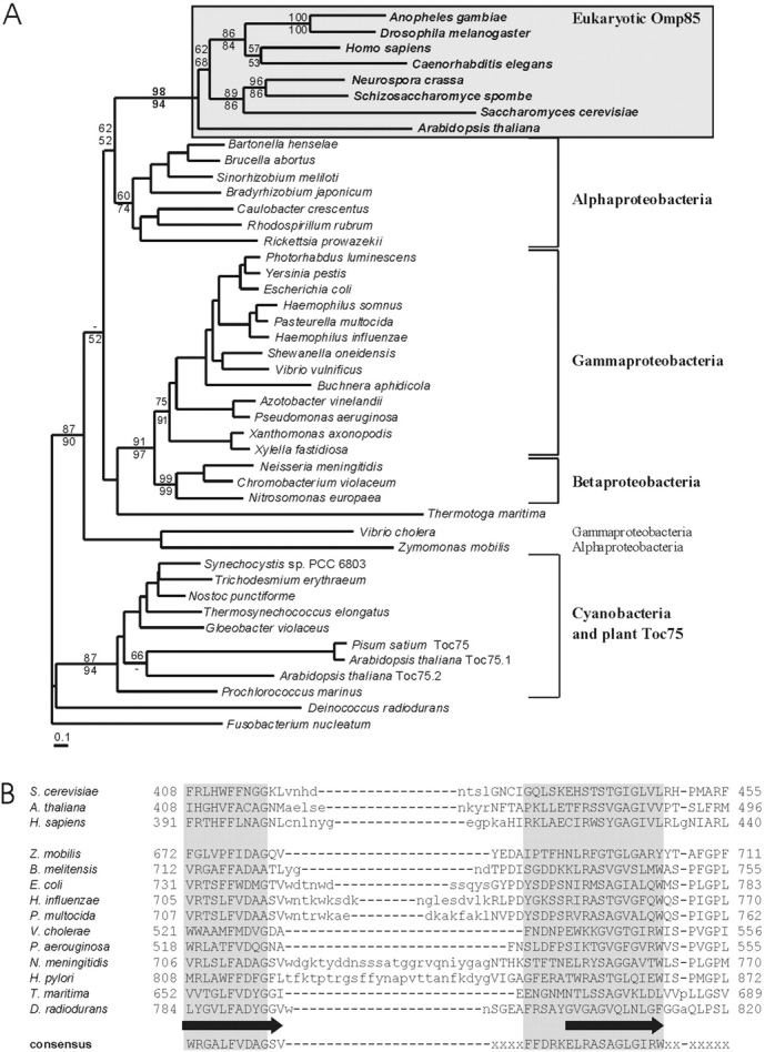
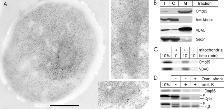
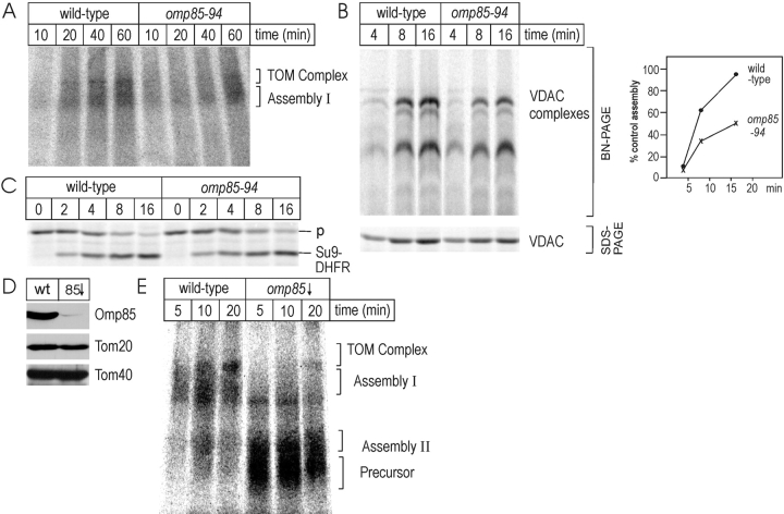
Integral proteins in the outer membrane of mitochondria control all aspects of organelle biogenesis, being required for protein import, mitochondrial fission, and, in metazoans, mitochondrial aspects of programmed cell death. How these integral proteins are assembled in the outer membrane had been unclear. In bacteria, Omp85 is an essential component of the protein insertion machinery, and we show that members of the Omp85 protein family are also found in eukaryotes ranging from plants to humans. In eukaryotes, Omp85 is present in the mitochondrial outer membrane. The gene encoding Omp85 is essential for cell viability in yeast, and conditional omp85 mutants have defects that arise from compromised insertion of integral proteins like voltage-dependent anion channel (VDAC) and components of the translocase in the outer membrane of mitochondria (TOM) complex into the mitochondrial outer membrane.


Figures

Figure 1.
Figure 1.
The Omp85 family. (A) Bootstrap support for eukaryote and other major nodes shown using weighted neighbor joining (top) and Fitch-Margoliash (bottom). Dashes indicate values <50%. (B) Sequence conservation between selected Omp85 family members and the consensus sequence derived with the conserved domain architecture retrieval tool (CDART; Geer et al., 2002). The most highly conserved section of the consensus sequence (shaded) includes the region corresponding to two predicted β-strands (designated by arrows), the least conserved (lower case) a predicted interstrand loop.
Figure 2.
Figure 2.
Omp85 is a mitochondrial outer membrane protein in S. cerevisiae. (A) Thin sections of high-pressure frozen, wild-type yeast were labeled with affinity-purified Omp85 antiserum and 15-nm colloidal gold. The entire cell (left) shows that labeling, though weak, is specific to the mitochondrial periphery. Four, two, and three gold particles are present in the left, upper right, and lower right images, respectively. Bars, 500 nm. (B) Yeast cells fractionated into total extracts (T), cytosol (C), and purified mitochondria (M) were analyzed by SDS-PAGE and immunoblotting for the mitochondrial protein VDAC, Sec61 from the membrane of the endoplasmic reticulum, and the cytosolic protein hexokinase. (C) 35S-labeled Omp85 and VDAC were synthesized in vitro (10% of the total shown) and imported. Mitochondria were then extracted with 0.1 M Na2CO3 and purified by flotation through a sucrose cushion containing 0.1 M Na2CO3. After SDS-PAGE, 35S-labeled Omp85 and VDAC were detected by fluorography. (D) Omp85, Cyb2, and F1β were synthesized in vitro and incubated with yeast mitochondria. Where shown, p indicates the precursor form of the proteins. Mitochondria were then treated with 50 μg/ml proteinase K (+) in iso-osmotic buffer or in hypo-osmotic buffer.
Figure 3.
Figure 3.
Omp85 mediates protein insertion and assembly into the mitochondrial outer membrane. (A) Mitochondria (50 mg) isolated from wild-type or mutant yeast cells were incubated with 35S-labeled Tom40 for the indicated times and then isolated for BN-PAGE and phosphorimage analysis. The identity of each TOM complex intermediate was estimated from the size of marker proteins (Model et al., 2001). (B) Mitochondria (50 mg) isolated from wild-type or mutant yeast cells were incubated with 35S-labeled VDAC. Duplicate assays were committed either to BN-PAGE or, after treatment with proteinase K, analyzed by SDS-PAGE and fluorography (Krimmer et al., 2001). Quantitation of the VDAC complexes is graphed beside the fluorogram. (C) 35S-labeled Su9-DHFR was incubated with mitochondria for the indicated times, and then the mitochondria were treated with proteinase K. Imported proteins were analyzed by SDS-PAGE and fluorography. p indicates the precursor form of Su9-DHFR. (D) Mitochondria were isolated from wild-type or omp85↓ cells and assayed by immunoblotting for levels of Omp85 and the TOM complex subunits Tom20 and Tom40. (E) Mitochondria (50 mg) isolated from wild-type or omp85↓ cells were incubated with 35S-labeled Tom40 for the indicated times and then isolated for BN-PAGE and phosphorimage analysis.

References

    1. Adams, J.M., and S. Cory. 2001. Life-or-death decisions by the Bcl-2 protein family. Trends Biochem. Sci. 26:61–66. - PubMed
    1. Bay, D.C., and D.A. Court. 2002. Origami in the outer membrane: the transmembrane arrangement of mitochondrial porins. Biochem. Cell Biol. 80:551–562. - PubMed
    1. Bruno, W.J., N.D. Socci, and A.L. Halpern. 2000. Weighted neighbor joining: a likelihood-based approach to distance-based phylogeny reconstruction. Mol. Biol. Evol. 17:189–197. - PubMed
    1. Buchanan, S.K. 1999. Beta-barrel proteins from bacterial outer membranes: structure, function and refolding. Curr. Opin. Struct. Biol. 9:455–461. - PubMed
    1. Cavalier-Smith, T. 2002. The phagotrophic origin of eukaryotes and phylogenetic classification of protozoa. Int. J. Syst. Evol. Microbiol. 52:297–354. - PubMed

Publication types

MeSH terms

Substances