Review
. 2004 Jan;229(1):143-61.
doi: 10.1002/dvdy.10444.

Turning heads: development of vertebrate branchiomotor neurons

Affiliations
Review

Turning heads: development of vertebrate branchiomotor neurons

Anand Chandrasekhar. Dev Dyn. 2004 Jan.

Abstract

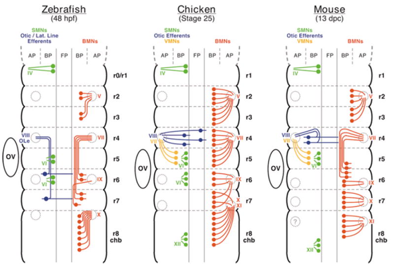
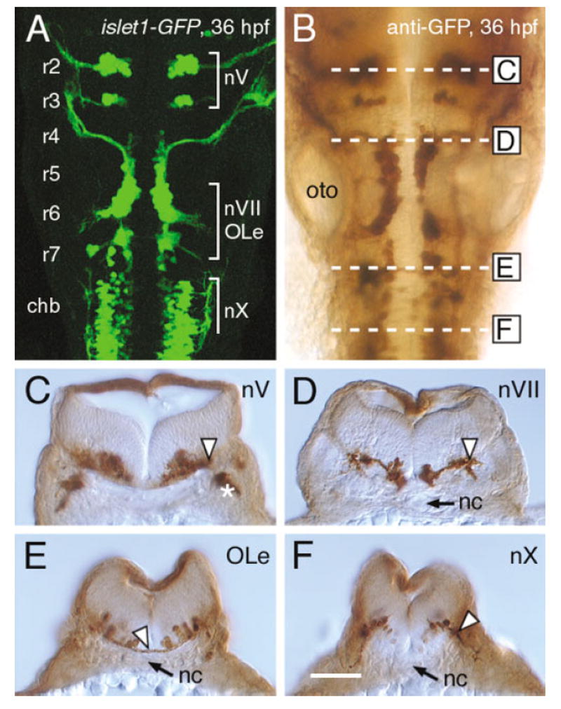
The cranial motor neurons innervate muscles that control eye, jaw, and facial movements of the vertebrate head and parasympathetic neurons that innervate certain glands and organs. These efferent neurons develop at characteristic locations in the brainstem, and their axons exit the neural tube in well-defined trajectories to innervate target tissues. This review is focused on a subset of cranial motor neurons called the branchiomotor neurons, which innervate muscles derived from the branchial (pharyngeal) arches. First, the organization of the branchiomotor pathways in zebrafish, chick, and mouse embryos will be compared, and the underlying axon guidance mechanisms will be addressed. Next, the molecular mechanisms that generate branchiomotor neurons and specify their identities will be discussed. Finally, the caudally directed or tangential migration of facial branchiomotor neurons will be examined. Given the advances in the characterization and analysis of vertebrate genomes, we can expect rapid progress in elucidating the cellular and molecular mechanisms underlying the development of these vital neuronal networks. Developmental Dynamics 229:143-161, 2004.


Figures

Fig. 1
Fig. 1
Organization of cranial motor neurons in zebrafish, chick, and mouse. Embryo ages: zebrafish, 48 hours postfertilization (hpf); chicken, stage 25; mouse, 13 days post coitum (dpc). The hindbrains are depicted in open-book configuration (anterior to the top), with the dorsal most neural tube at the lateral margins of the diagrams. The series of bulges and the broken grey lines partition the hindbrain into rhombomeres from r0/r1 to r8. The hindbrain region anterior to r2 is defined as r0/r1, because it is composed of two distinct domains, r0 or the isthmic rhombomere anteriorly, and r1 posteriorly, which can be distinguished using molecular markers in zebrafish (Moens and Prince, 2002). Rhombomere 8 is longer than the other rhombomeres, and is also referred to as the caudal hindbrain (chb). The branchiomotor neurons (BMNs) and axons are shown in red, the somatomotor neurons (SMNs) in green, the otic and lateral line efferent (VIII, OLe) neurons in blue, and the visceromotor neurons (VMNs) in yellow (see text for details). For clarity, motor neuron populations are depicted on only one side of the neural tube. In the chick and mouse neural tubes, the alar plate (AP) and basal plate (BP) are clearly demarcated by the sulcus limitans. The zebrafish neural tube forms through different morphogenetic movements compared with chick and mouse, and the distinction between AP and BP in the hindbrain has not been described. Therefore, the AP and BP assignments are tentative in the fish neural tube. FP marks the floor plate region. The nerve exit points of the BMN, VMN, and otic efferent axons are shown as grey circles and are located dorsally in the alar plate of the chick and mouse neural tubes. In contrast, the exit points appear to be more ventral in zebrafish, and their depicted locations relative to the AP–BP boundary are tentative. The exit points of the SMN axons are located ventrally within the BP, close to the FP, in all species. The uncertainty in the location of the nerve XI exit point in the mouse embryo is indicated by a question mark. OV, otic vesicle.
Fig. 2
Fig. 2
Location of the cranial nerve exit points in the zebrafish hindbrain. A,B: Dorsal views of the hindbrain, with anterior to the top, in 36 hours postfertilization (hpf) transgenic embryos expressing green fluorescent protein (GFP) in the trigeminal motor (nV), facial motor (nVII), otic and lateral line efferent (OLe), and vagal motor (nX) neurons. GFP was detected by confocal imaging (A), and by labeling with an anti-GFP antibody (B). The various motor and efferent neuron populations are found at characteristic locations in rhombomeres 2–7 (r2–r7), and the caudal hindbrain (chb) (see text for details). The broken white lines in B indicate the plane of the cross-sections depicted in C–F. oto, otic vesicle. C–F: Cross-sections of the hindbrain in GFP antibody-labeled embryos at the approximate axial levels indicated in B. Dorsal is to the top in all panels. nc, notochord. C: The nV branchiomotor neuron cell bodies in r2 are located medially, close to the floor plate, and the exit point for their axons is located dorsolaterally (arrowhead). The asterisk indicates the nVII motor axons extending into the second branchial arch. D: Many nVII/OLe cell bodies are still found in r4 in this embryo, immediately adjacent to the floor plate. The axons are tightly fasciculated and exit the hindbrain dorsolaterally (arrowhead). E: In r7, GFP-labeled processes of nVII/OLe neurons (arrowhead) cross the midline ventral to the floor plate cells. Based on their similarity to the projections of contralateral vestibuloacous-tic neurons in chick and mouse, these processes are tentatively assigned to zebrafish OLe neurons (see text for details). F: The nX motor neurons are located more dorsolaterally compared with the nV and nVII neurons, and their axons exit from the neural tube (arrowhead) where the cell bodies are located. Scale bar in F = 40 μm in A,B, 30 μm in C–F.

Similar articles

Cited by

References

    1. Abadie V, Champagnat J, Fortin G. Branchiomotor activities in mouse embryo. Neuroreport. 2000;11:141–145. - PubMed
    1. Abadie V, Morisseau-Durand MP, Beyler C, Manach Y, Couly G. Brainstem dysfunction: a possible neuroembryological pathogenesis of isolated Pierre Robin sequence. Eur J Pediatr. 2002;161:275–280. - PubMed
    1. Ando H, Furuta T, Tsien RY, Okamoto H. Photo-mediated gene activation using caged RNA/DNA in zebrafish embryos. Nat Genet. 2001;28:317–325. - PubMed
    1. Appel B, Korzh V, Glasgow E, Thor S, Edlund T, Dawid IB, Eisen JS. Motoneuron fate specification revealed by patterned LIM homeobox gene expression in embryonic zebrafish. Development. 1995;121:4117–4125. - PubMed
    1. Barrett C, Guthrie S. Expression patterns of the netrin receptor UNC5H1 among developing motor neurons in the embryonic rat hindbrain. Mech Dev. 2001;106:163–166. - PubMed

LinkOut - more resources