Review
. 2004 Jan;276(1):2-12.
doi: 10.1002/ar.a.10130.

Stem cells and the formation of the myocardium in the vertebrate embryo

Affiliations
Review

Stem cells and the formation of the myocardium in the vertebrate embryo

Leonard M Eisenberg et al. Anat Rec A Discov Mol Cell Evol Biol. 2004 Jan.

Abstract

A major goal in cardiovascular biology is to repair diseased or damaged hearts with newly generated myocardial tissue. Stem cells offer a potential source of replacement myocytes for restoring cardiac function. Yet little is known about the nature of the cells that are able to generate myocardium and the conditions they require to form heart tissue. A source of information that may be pertinent to addressing these issues is the study of how the myocardium arises from progenitor cells in the early vertebrate embryo. Accordingly, this review will examine the initial events of cardiac developmental biology for insights into the identity and characteristics of the stem cells that can be used to generate myocardial tissue for therapeutic purposes.


Figures

Fig. 1
Fig. 1
Schematic diagram depicting the initial morphological events in the development of the avian heart. Precardiac and definitive myocardial tissue are illustrated in black, while the lines overlaying the cardiac areas denote the plane of the transverse sections shown immediately below the embryos. A: Three-dimensional view of a cross-sectioned chick embryo, which is in transition between HH stages 3 and 4. Precardiac cells undergo gastrulation through the primitive streak and move laterally to reside in lateral mesoderm within the anterior half of the embryo. B: During HH stage 5, the myocardial progenitors coalesce into morphologically distinct heart-forming fields, which are distributed bilaterally to the primitive streak. C: At HH stage 8, the two heart-forming areas have sorted to splanchnic mesoderm and begun to merge at the embryonic midline. By this stage, these cells have become definitive cardiac myocytes, as they will exhibit a number of muscle proteins in a prestriated pattern. D: By HH stage 10 a fully contractile heart has developed. This primitive heart tube consists of an outer sheet of myocardium, surrounding an inner endocardial layer.
Fig. 2
Fig. 2
Location of precardiac cells for HH stages 3–5, as determined by the fate map studies of Stalsberg and DeHaan (1969), Garcia-Martinez and Schoenwolf (1993), and Redkar et al. (2001). The precardiac cells are exhibited within the caudal half of the primitive streak in HH stage 3 avian embryo. At HH stages 4 and 5, precardiac cells become distributed bilaterally and reside within the anterior lateral mesoderm. Precardiac areas are shown in gray.
Fig. 3
Fig. 3
Multipotentiality of precardiac mesoderm. A and B: Precardiac mesoderm possesses hematopoietic potential. Precardiac mesoderm from HH stage 5 (A) and HH stage 6 (B) mesodermal cells obtained from precardiac regions were dissociated and cultured for 4 days in fibrin gels under conditions that promote hematopoiesis (Brandon et al., 2000). The presence of blood cells was verified by cytological examination with May-Grünwald-Giemsa stain. Panels A and B display clusters of monocytes and macrophages, respectively. C–I: Multipotentiality of the precardiac mesoderm-derived cell line QCE6. C: QCE6 cells undergo myocardial differentiation when co-cultured with HH stage 15 embryonic chick heart tissue. After 48 hr of incubation, the cultures were dual stained for both sarcomeric myosin (green) and β-galactosidase (a marker of the virally labeled QCE6 cells, red). The green staining on the right is sarcomeric myosin-positive ventricular tissue, while in the center of the field are QCE6 cells that incorporated into the heart tissue and thus subsequently exhibited a cardiac phenotype. This is indicated by the dual reactivity with both antibodies, which produced the yellow staining (arrow). D–F: Endothelial differentiation of QCE6 cells as indicated by branching morphogenesis and von Willebrand factor (vWF) expression. Cells were treated for 48 hr, immunostained for vWF, and imaged by brightfield (D), phase (E), and fluorescent microscopy (F). Low- (D) and high-magnification (E) views show extensive branching morphogenesis. F: Moreover, these cultures produced high levels of vWF protein. Arrow in panels D–F corresponds to the identical position within the culture. G–I: Hematopoiesis of QCE6 cells. Cells were cultured as described previously (Brandon et al., 2000) and stained with May-Grünwald-Giemsa dye to identify specific blood cell phenotypes. Here are shown QCE6-derived monocytes (G), macrophages (H), and red blood cells (I).
Fig. 4
Fig. 4
Myocardial potential of early avian mesoderm. Explants containing lateral mesoderm and the underlying endoderm were microdissected from HH stage 4, 5, and 6 avian embryos and cultured in minimal media. Subsequently, the presence of cardiac tissue was verified by immunostaining cultures for sarcomeric myosin. As previously described (Eisenberg and Eisenberg, 1999), explants containing precardiac mesoderm from each of the stages readily developed into myocardial tissue. In contrast, tissue from posterior noncardiac regions of HH stage 5 or stage 6 did not undergo cardiac differentiation in culture. Surprisingly, posterior lateral mesoderm from HH stage 4 embryos, which does not contribute to the heart in the embryo, generated cardiac tissue in culture. The total number of explants examined for each group is listed above each bar.
Fig. 5
Fig. 5
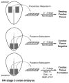
The formation of myocardial tissue from embryonic avian explants. As depicted in this schematic diagram, tissue containing lateral mesoderm and the underlying endoderm (mesendoderm) from either anterior precardiac or posterior noncardiac regions was removed from HH stage 5 embryos and cultured for cultured 2–3 days. Tissue obtained from anterior precardiac areas will form beating tissue within the first 30 hr of culture and display large regions of myocardium. In contrast, explants of noncardiac posterior mesendoderm will not generate cardiac tissue. However, treatment of noncardiac posterior mesendoderm with WNT11 (Eisenberg and Eisenberg, 1999), Dkk1 (Marvin et al., 2001), or BMP4 + FGF2 (Lough et al., 1996; Ladd et al., 1998) will promote the development of cardiac tissue, as exhibited by clustered fields of cells expressing cardiac proteins. Thus, despite the posterior origin of these latter explants, which correspond to regions of the embryo that do not contribute to the heart in situ, this tissue is still able to form myocardial tissue in response to changes in the extracellular environment.
Fig. 6
Fig. 6
Incorporation and differentiation of circulating cells into the embryonic myocardium. ED9 mouse embryos were dissected from the uterus with fetal placenta and yolk sac intact and then injected into the yolk sac vessel leading to the sinus venous with QCE6 cells labeled with either a β-galactosidase-positive retrovirus (Eisenberg and Markwald, 1997) (A–C) or the fluorescent cell marker DiI (1,1′-dioctadecyl-3, 3,3′, 3′-tetramethyl-indocarbocyanine perchlorate) (D–F). The injected embryos were allowed to develop for 24 hr ex ovo, according to established procedures (Sturm and Tam, 1993; Eto and Osumi-Yamashita, 1995). A–C: QCE6 cells in the outer myocardial wall overlying the AV canal. X-gal staining for β-galactosidase (blue) indicates the presence of QCE6 cells (arrow) at low(A) and high (B) magnification. C: A sister section stained with the MF20 antibody, which recognizes sarcomeric myosin heavy chain (sMyHC). The arrow indicates a sMyHC-positive QCE6 cell and thus displays a myocardial phenotype. D–F: A DiI-labeled QCE6 cell in the myocardium of the outflow tract. An individual heart section is exhibited for DiI-labeling only (red) (D), sMyHC immunostaining only (green) (E), or both DiI labeling and sMyHC staining (F). The juxtaposition of these panels demonstrates that the DiI-labeled QCE6 cell (arrow) is sMyHC positive. The myocardial (myo) and endocardial (endo) layers of the heart tissue are indicated in panel D.

References

    1. Alsan BH, Schultheiss TM. Regulation of avian cardiogenesis by Fgf8 signaling. Development. 2002;129:1935–1943. - PubMed
    1. Antin PB, Taylor RG, Yatskievych T. Precardiac mesoderm is specified during gastrulation in quail. Dev Dyn. 1994;200:144–154. - PubMed
    1. Anversa P, Nadal-Ginard B. Myocyte renewal and ventricular remodelling. Nature. 2002;415:240–243. - PubMed
    1. Beltrami AP, Urbanek K, Kajstura J, Yan S-M, Finato N, Bussani R, Nadal-Ginard B, Silvestri F, Leri A, Beltrami CA, Anversa P. Evidence that human cardiac myocytes divide after myocardial infarction. N Engl J Med. 2001;344:1750–1757. - PubMed
    1. Biben C, Hatzistavrou T, Harvey RP. Expression of NK-2 class homeobox gene Nkx2-6 in foregut endoderm and heart. Mech Dev. 1998;73:125–127. - PubMed

Publication types

LinkOut - more resources