. 2004 Jan;24(2):514-26.
doi: 10.1128/MCB.24.2.514-526.2004.

Mitotic degradation of human thymidine kinase 1 is dependent on the anaphase-promoting complex/cyclosome-CDH1-mediated pathway

Affiliations

Mitotic degradation of human thymidine kinase 1 is dependent on the anaphase-promoting complex/cyclosome-CDH1-mediated pathway

Po-Yuan Ke et al. Mol Cell Biol. 2004 Jan.

Abstract

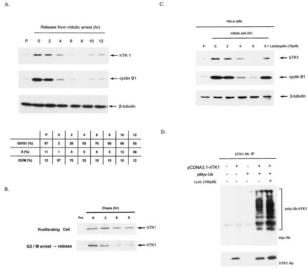
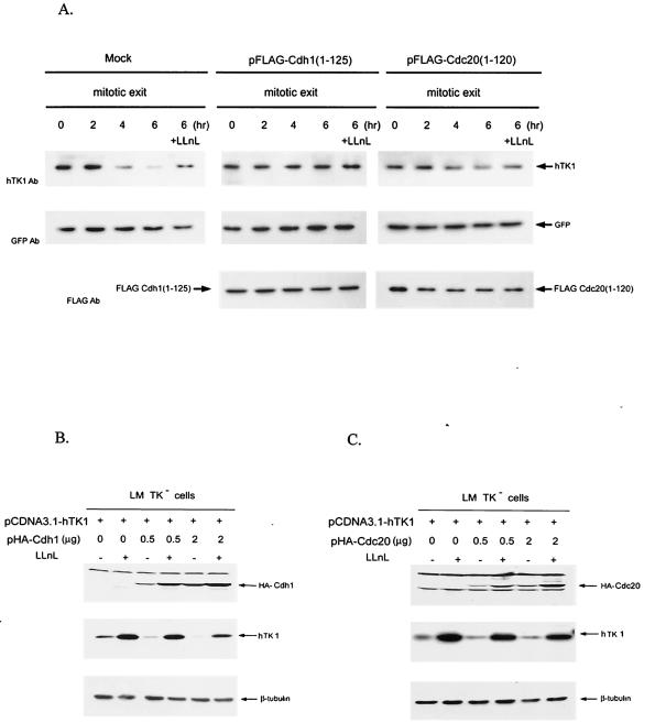
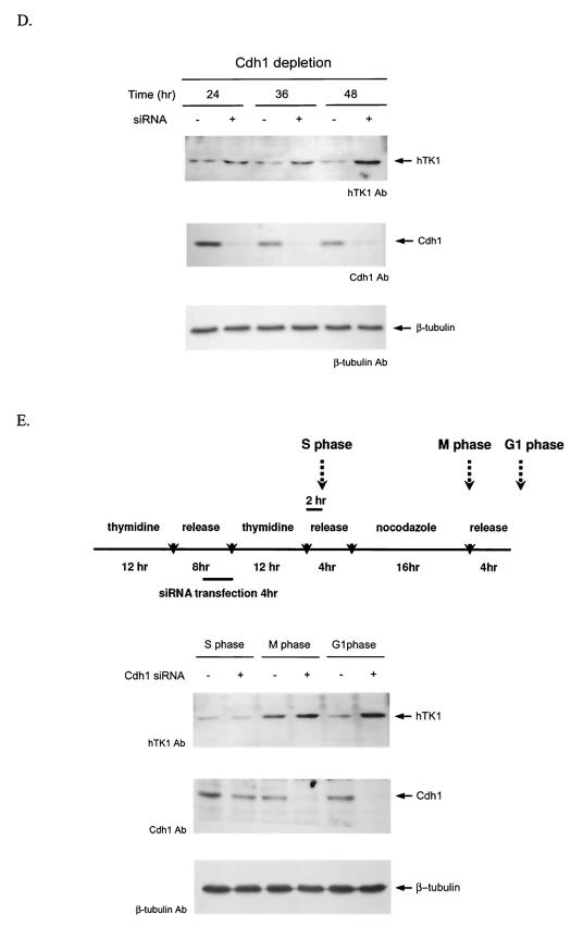
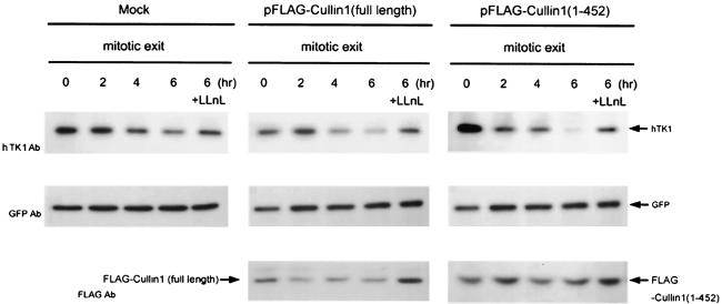
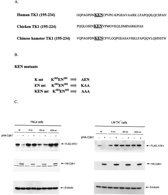
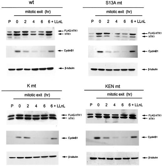
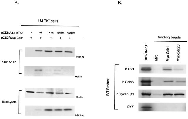
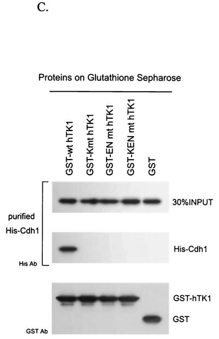
The expression of human thymidine kinase 1 (hTK1) is highly dependent on the growth states and cell cycle stages in mammalian cells. The amount of hTK1 is significantly increased in the cells during progression to the S and M phases, and becomes barely detectable in the early G(1) phase by a proteolytic control during mitotic exit. This tight regulation is important for providing the correct pool of dTTP for DNA synthesis at the right time in the cell cycle. Here, we investigated the mechanism responsible for mitotic degradation of hTK1. We show that hTK1 is degraded via a ubiquitin-proteasome pathway in mammalian cells and that anaphase-promoting complex/cyclosome (APC/C) activator Cdh1 is not only a necessary but also a rate-limiting factor for mitotic degradation of hTK1. Furthermore, a KEN box sequence located in the C-terminal region of hTK1 is required for its mitotic degradation and interaction capability with Cdh1. By in vitro ubiquitinylation assays, we demonstrated that hTK1 is targeted for degradation by the APC/C-Cdh1 ubiquitin ligase dependent on this KEN box motif. Taken together, we concluded that activation of the APC/C-Cdh1 complex during mitotic exit controls timing of hTK1 destruction, thus effectively minimizing dTTP formation from the salvage pathway in the early G(1) phase of the cell cycle in mammalian cells.


Figures

FIG. 1.
FIG. 1.
Mitotic degradation of hTK1 is via the ubiquitin-proteasomal pathway. (A) HeLa cells were arrested by nocodazole treatment for 20 h, followed by replenishment of fresh medium to release cells from the G2/M arrest. Extracts were prepared from HeLa cells released from G2/M arrest at the time points as indicated. The extract from asynchronized cells is indicated as the proliferating state (P). Expression of hTK1, cyclin B1, and β-tubulin was analyzed by Western blotting with anti-hTK1, anti-cyclin B1, and anti-β-tubulin antibodies, respectively. The cell cycle profile of corresponding cells was analyzed by flow cytometry as described in Materials and Methods and is expressed in the inset tables as percentages of cells in different phases. (B) HeLa cells of proliferating state or releasing from G2/M arrest were metabolically labeled with [35S]methionine (250 μCi/ml) for 30 min, followed by addition of fresh medium at the indicated time points prior to cell lysis. Cell extracts were immunoprecipitated by anti-hTK1 antibody, and the immunocomplexes were resolved by SDS-PAGE (12% polyacrylamide), followed by autoradiography. Normal rabbit immunoglobulin G (Pre) was used as a negative control. (C) Extracts were prepared from HeLa cells released from G2/M arrest. Cells were treated with lactacystin (10 μM) for 6 h during the mitotic exit phase. (D) LM-TK cells were transfected with (+) or without (−) pCDNA3.1-hTK1 and pMyc-Ub as indicated. hTK1 was immunoprecipitated (IP) with anti-hTK1 antibody (Ab) and immunocomplexes were analyzed by SDS-PAGE and Western blotting with anti-hTK1 and anti-Myc antibodies.
FIG. 2.
FIG. 2.
Mitotic degradation of hTK1 requires functional Cdh1. (A) HeLa cells were transfected with pFLAG-Cdc20(1-120), pFLAG-Cdh1(1-125) or empty vector (Mock) together with pEGFP as an internal control. Following transfection for 24 h, cells were synchronized as described in the legend to Fig. 1. One set of cells during release from the mitotic arrest was treated with LLnL (100 μM) for 6 h. Expression of hTK1, Cdc20(1-120), Cdh1(1-125), and GFP was analyzed by Western blotting with specific antibodies (Ab). (B) LM TK cells were transfected with 1 μg of pCDNA3.1-hTK1 and different amounts of pHA-Cdh1 as indicated. Empty vector was added to have a final 3 μg of total DNA for transfection. Cells were treated without (−) or with (+) LLnL (100 μM) for 6 h before harvesting. HA-Cdh1, hTK1, and β-tubulin were detected by Western blot analysis. (C) LM-TK− cells transfected with pCDNA3.1-hTK1 and different amounts of pHA-Cdc20 as indicated were analyzed as described above. (D) HeLa cells were transfected without (−) or with (+) 0.8 μg of duplex siRNA against Cdh1 as described in Materials and Methods. After transfection at different time points, cells were extracted for Western blot analysis. (E) Scheme of the synchronization procedures for Cdh1 siRNA transfection experiment in HeLa cells (upper panel). Cell extracts were prepared after transfection at different phases as indicated and analyzed as described above.
FIG. 2.
FIG. 2.
Mitotic degradation of hTK1 requires functional Cdh1. (A) HeLa cells were transfected with pFLAG-Cdc20(1-120), pFLAG-Cdh1(1-125) or empty vector (Mock) together with pEGFP as an internal control. Following transfection for 24 h, cells were synchronized as described in the legend to Fig. 1. One set of cells during release from the mitotic arrest was treated with LLnL (100 μM) for 6 h. Expression of hTK1, Cdc20(1-120), Cdh1(1-125), and GFP was analyzed by Western blotting with specific antibodies (Ab). (B) LM TK cells were transfected with 1 μg of pCDNA3.1-hTK1 and different amounts of pHA-Cdh1 as indicated. Empty vector was added to have a final 3 μg of total DNA for transfection. Cells were treated without (−) or with (+) LLnL (100 μM) for 6 h before harvesting. HA-Cdh1, hTK1, and β-tubulin were detected by Western blot analysis. (C) LM-TK− cells transfected with pCDNA3.1-hTK1 and different amounts of pHA-Cdc20 as indicated were analyzed as described above. (D) HeLa cells were transfected without (−) or with (+) 0.8 μg of duplex siRNA against Cdh1 as described in Materials and Methods. After transfection at different time points, cells were extracted for Western blot analysis. (E) Scheme of the synchronization procedures for Cdh1 siRNA transfection experiment in HeLa cells (upper panel). Cell extracts were prepared after transfection at different phases as indicated and analyzed as described above.
FIG. 3.
FIG. 3.
The function of SCF is not essential for mitotic degradation of hTK1. HeLa cells were transfected with pFLAG-Cullin1(full length), pFLAG-Cullin1(1-452), or empty vector (Mock) together with pEGFP as an internal control. Cell synchronization, LLnL treatment, and analysis of protein abundance were performed as described in the legend to Fig. 2.
FIG. 4.
FIG. 4.
Cdh1-dependent degradation of hTK1 requires the KEN box motif located at the C-terminal region. (A) Sequence alignment of the C-terminal region covering aa 195 to 234 of hTK1 with the corresponding regions of other species. The KEN box is indicated in boldface. (B) Schematic representation of mutants carrying a mutation in the KEN box sequence. (C) Extracts were prepared from HeLa cells (left panel) and LM-TK cells (right panel) transfected with pFLAG-hTK1 (wild type [wt]) or KEN box mutants (K mt, EN mt, and KEN mt) with (+) or without (−) pHA-Cdh1, followed by SDS-PAGE separation and Western blot analysis as described in the legend to Fig. 2B.
FIG. 5.
FIG. 5.
The KEN box is a necessary signal for mitotic degradation of hTK1. Stable cell lines expressing the wild type (wt), Ser13A-mutated (S13A mt), and KEN box-mutated (K mt and KEN mt) FLAG-hTK1 were prepared as described in Materials and Methods. Mitotic arrest of cells and analysis of protein abundance were performed as described in the legend to Fig. 1A.
FIG. 6.
FIG. 6.
hTK1 interacts with Cdh1 via the KEN box. (A) LM-TK cells were transfected with pCDNA3.1-hTK1 of the wild type (wt) or KEN box mutants (K mt, EN mt, and KEN mt) together with pCS2+Myc-Cdh1. After 24 h, cells were treated with LLnL (100 μM) for 6 h before harvesting. hTK1 was immunoprecipitated as described in the legend to Fig. 1D and analyzed by Western blotting with specific antibodies. (B) In vitro-translated 35S-labeled hTK1, hCdc6, human cyclin B1, and p27 were individually incubated with anti-Myc antibody-coated beads containing the unlabeled in vitro-translated Myc-Cdh1, Myc-Cdc20, or Myc. The left lane indicates 10% of the 35S-labeled in vitro-translated product in each binding assay. (C) Purified His-Cdh1 proteins were subjected into GST pull-down assays using glutathione-Sepharose beads bound with GST, GST-hTK1 (wild type), and three KEN box mutants as described in Materials and Methods. The pull-down samples were analyzed by SDS-PAGE and Western blotting with specific antibodies.
FIG. 6.
FIG. 6.
hTK1 interacts with Cdh1 via the KEN box. (A) LM-TK cells were transfected with pCDNA3.1-hTK1 of the wild type (wt) or KEN box mutants (K mt, EN mt, and KEN mt) together with pCS2+Myc-Cdh1. After 24 h, cells were treated with LLnL (100 μM) for 6 h before harvesting. hTK1 was immunoprecipitated as described in the legend to Fig. 1D and analyzed by Western blotting with specific antibodies. (B) In vitro-translated 35S-labeled hTK1, hCdc6, human cyclin B1, and p27 were individually incubated with anti-Myc antibody-coated beads containing the unlabeled in vitro-translated Myc-Cdh1, Myc-Cdc20, or Myc. The left lane indicates 10% of the 35S-labeled in vitro-translated product in each binding assay. (C) Purified His-Cdh1 proteins were subjected into GST pull-down assays using glutathione-Sepharose beads bound with GST, GST-hTK1 (wild type), and three KEN box mutants as described in Materials and Methods. The pull-down samples were analyzed by SDS-PAGE and Western blotting with specific antibodies.
FIG. 7.
FIG. 7.
APC/C-Cdh1 is the direct ubiquitin E3 ligase confers polyubiquitinylation of hTK1. (A) The early G1-phase extracts after depletion by control mouse immunoglobulin G (IgG), Cdc27, and Skp1 antibodies as described in Materials and Methods (representing complete, APC/C-negative, and SCF-negative extract, respectively) were subjected to Western blotting analysis with anti-Cdc27, anti-Skp1, and anti-β-tubulin antibodies. (B) An in vitro ubiquitinylation reaction was carried out with in vitro-translated 35S-labeled hTK1 using the complete, SCF-depleted (−SCF), or APC-depleted (−APC) extracts for the time indicated. The reaction products were analyzed by SDS-PAGE (9% polyacrylamide) prior to autoradiography. The hTK1-(Ub)1, hTK1-(Ub)2, and poly-Ub-hTK1 indicate the mono-, di-, and polyubiquitinylation of hTK1. (C) Reconstitution of in vitro ubiquitinylation of hTK1 was performed with His-hTK1 (wild type) or KEN mutant as a substrate in each reaction mixture containing E1, E2 (UbcX), APC/C, and Cdc20 or Cdh1 as indicated. The samples were analyzed by SDS-PAGE (5 to 15% polyacrylamide) and Western blotting with anti-hTK1 antibody.
FIG. 7.
FIG. 7.
APC/C-Cdh1 is the direct ubiquitin E3 ligase confers polyubiquitinylation of hTK1. (A) The early G1-phase extracts after depletion by control mouse immunoglobulin G (IgG), Cdc27, and Skp1 antibodies as described in Materials and Methods (representing complete, APC/C-negative, and SCF-negative extract, respectively) were subjected to Western blotting analysis with anti-Cdc27, anti-Skp1, and anti-β-tubulin antibodies. (B) An in vitro ubiquitinylation reaction was carried out with in vitro-translated 35S-labeled hTK1 using the complete, SCF-depleted (−SCF), or APC-depleted (−APC) extracts for the time indicated. The reaction products were analyzed by SDS-PAGE (9% polyacrylamide) prior to autoradiography. The hTK1-(Ub)1, hTK1-(Ub)2, and poly-Ub-hTK1 indicate the mono-, di-, and polyubiquitinylation of hTK1. (C) Reconstitution of in vitro ubiquitinylation of hTK1 was performed with His-hTK1 (wild type) or KEN mutant as a substrate in each reaction mixture containing E1, E2 (UbcX), APC/C, and Cdc20 or Cdh1 as indicated. The samples were analyzed by SDS-PAGE (5 to 15% polyacrylamide) and Western blotting with anti-hTK1 antibody.

References

    1. Bello, L. J. 1974. Regulation of thymidine kinase synthesis in human cells. Exp. Cell Res. 89:263-274. - PubMed
    1. Bradshaw, H. D., Jr., and P. L. Deininger. 1984. Human thymidine kinase gene: molecular cloning and nucleotide sequence of a cDNA expressible in mammalian cells. Mol. Cell. Biol. 4:2316-2320. - PMC - PubMed
    1. Brandeis, M., and T. Hunt. 1996. The proteolysis of mitotic cyclins in mammalian cells persists from the end of mitosis until the onset of S phase. EMBO J. 15:5280-5289. - PMC - PubMed
    1. Chabes, A. L., C. M. Pfleger, M. W. Kirschner, and L. Thelander. 2003. Mouse ribonuclotide reductase R2 protein: a new target for anaphase-promoting complex-Cdh1-mediated proteolysis. Proc. Natl. Acad. Sci. USA 100:3925-3929. - PMC - PubMed
    1. Chang, Z. F., D. Y. Huang, and N. C. Hsue. 1994. Differential phosphorylation of human thymidine kinase in proliferating and M phase-arrested human cells. J. Biol. Chem. 269:21249-21254. - PubMed

Publication types

MeSH terms

LinkOut - more resources