. 2004 Jan;24(2):550-60.
doi: 10.1128/MCB.24.2.550-560.2004.

Early glomerular filtration defect and severe renal disease in podocin-deficient mice

Affiliations

Early glomerular filtration defect and severe renal disease in podocin-deficient mice

Séverine Roselli et al. Mol Cell Biol. 2004 Jan.

Abstract

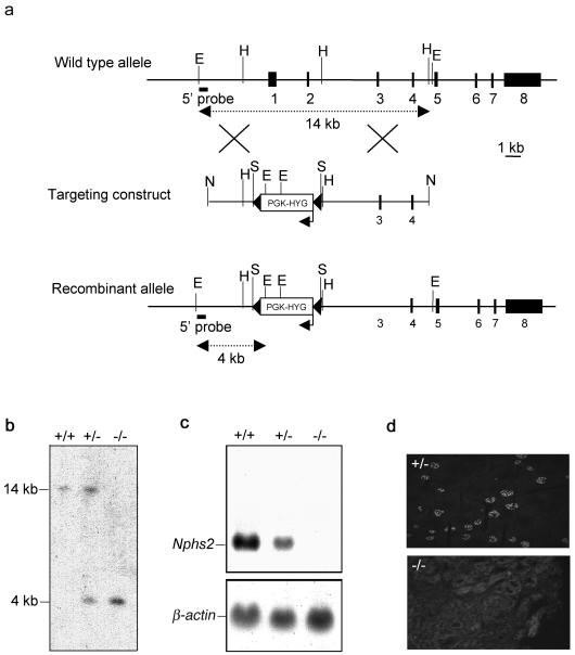
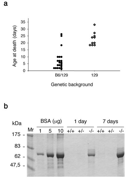
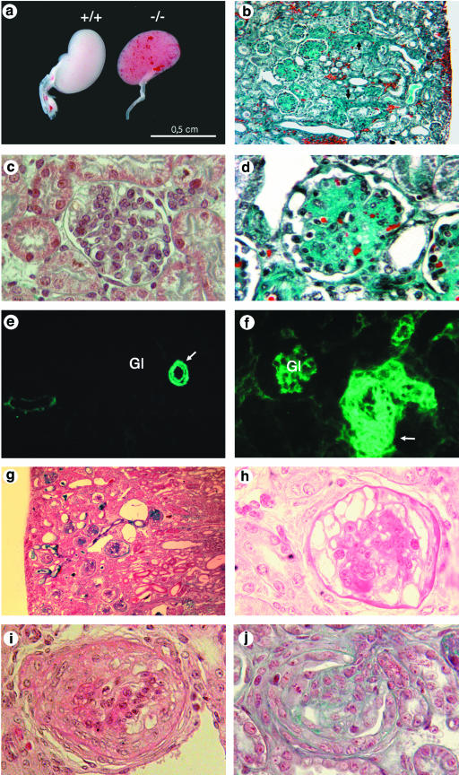
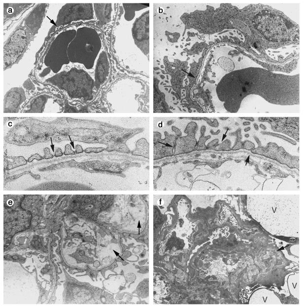
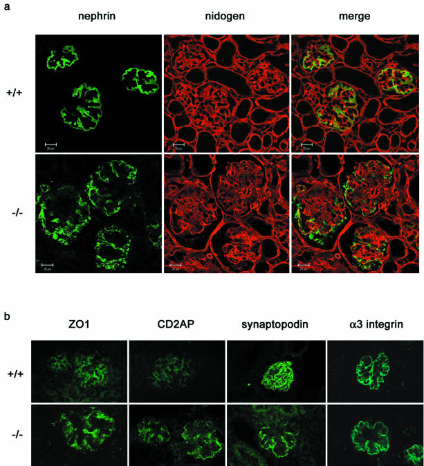
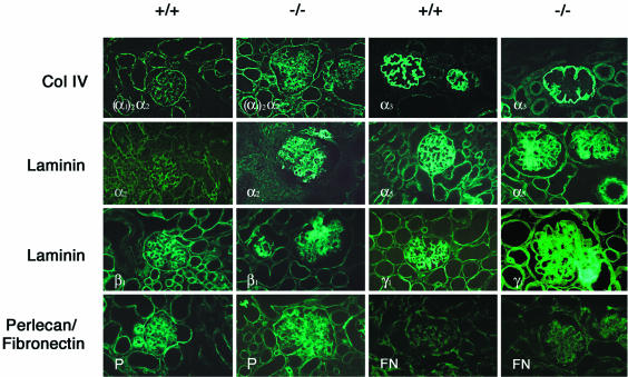
Podocytes are specialized epithelial cells covering the basement membrane of the glomerulus in the kidney. The molecular mechanisms underlying the role of podocytes in glomerular filtration are still largely unknown. We generated podocin-deficient (Nphs2-/-) mice to investigate the function of podocin, a protein expressed at the insertion of the slit diaphragm in podocytes and defective in a subset of patients with steroid-resistant nephrotic syndrome and focal and segmental glomerulosclerosis. Nphs2-/- mice developed proteinuria during the antenatal period and died a few days after birth from renal failure caused by massive mesangial sclerosis. Electron microscopy revealed the extensive fusion of podocyte foot processes and the lack of a slit diaphragm in the remaining foot process junctions. Using real-time PCR and immunolabeling, we showed that the expression of other slit diaphragm components was modified in Nphs2-/- kidneys: the expression of the nephrin gene was downregulated, whereas that of the ZO1 and CD2AP genes appeared to be upregulated. Interestingly, the progression of the renal disease, as well as the presence or absence of renal vascular lesions, depends on the genetic background. Our data demonstrate the crucial role of podocin in the establishment of the glomerular filtration barrier and provide a suitable model for mapping and identifying modifier genes involved in glomerular diseases caused by podocyte injuries.


Figures

FIG. 1.
FIG. 1.
Disruption of the Nphs2 gene. (a) Schematic representation of the genomic structure of the wild-type murine Nphs2 allele (top), the targeting construct (middle), and the targeted allele (bottom). The eight exons are represented by closed boxes. The region containing exons 1 and 2 was replaced by a floxed PGK-hygromycin cassette (PGK-HYG) (the arrow indicates the orientation of this gene). The LoxP sites are represented by black arrowheads. Restriction enzyme cleavage sites are indicated (E, EcoRI; H, HindIII, S, SmaI, N, NotI). (b) Southern blot analysis of genomic DNA from Nphs2+/+, Nphs2+/−, and Nphs2−/− mice with a 5′-external probe (solid bar). The 14-kb wild-type EcoRI fragment was detected in the Nphs2+/+ mouse; the recombinant 4-kb fragment, created due to the presence of an EcoRI site in the cassette, was detected in the Nphs2−/− mouse, and both bands were detected in the Nphs2+/− mouse. (c) Northern blot analysis of 20 μg of total kidney RNA from Nphs2+/+, Nphs2+/−, and Nphs2−/− mice hybridized with a 3′ cDNA probe spanning exons 4 to 8. The endogenous 3.1-kb Nphs2 transcript was detected in Nphs2+/+ mice, no transcript was detected in Nphs2−/− mice due to the deletion of the transcription start site, and the endogenous 3.1-kb transcript was detected in Nphs2+/− mice, although the band was weaker than in Nphs2+/+ mice. (d) Immunofluorescence study on cryostat sections of kidneys from Nphs2+/− and Nphs2−/− mice with a rabbit anti-human antibody directed against the C-terminal part of podocin. Podocin was detected in the glomeruli of Nphs2+/− mice, whereas no signal was observed in the Nphs2−/− kidney, a finding consistent with the lack of Nphs2 transcripts.
FIG. 2.
FIG. 2.
Clinical features of Nphs2−/− mice. (a) Schematic representation of the survival of Nphs2−/− mice from the mixed B6/129 and the pure 129 genetic backgrounds; (b) SDS-PAGE analysis of urine from Nphs2+/+, Nphs2+/−, and Nphs2−/− B6/129 littermates (1 and 7 days old). One microliter of urine from each mouse and albumin standards were loaded on the same gel. Massive proteinuria, mainly consisting of albumin, was observed in Nphs2−/− mice from birth but not in control mice.
FIG.3.
FIG.3.
Histologic analysis of Nphs2−/− mice. (a to f) Mixed B6/129 genetic background. (a) Kidneys from Nphs2+/+ and Nphs2−/− mice at 3 days of age. Red spots due to localized hemorrhages are visible at the surface of the Nphs2−/− mouse kidneys. (b) Diffuse glomerular involvement in a 7-day-old mouse associated with focal tubular dilatation, arteriolar thickening (arrow), and interstitial hemorrhages (magnification, ×180). (c) Normal mouse glomerulus (magnification, ×800). (d) Massive mesangial sclerosis in a 7-day-old mouse (magnification, ×720). (e and f) Immunohistochemical analysis of αSMA distribution. (e) In a 6-day-old wild-type mouse kidneys, αSMA labeling is strong in arterial and arteriolar smooth muscle cells (arrow) and negative in mature glomeruli (Gl). (f) In a 6-day-old Nphs2−/− mouse, marked thickening of the arterial wall is accentuated by the presence of several layers of αSMA-positive cells (arrow). In addition, high levels of αSMA were observed in mesangial cells of the glomeruli (Gl). (g and h) Collapsing form of glomerulopathy in one 24-day-old Nphs2−/− mouse of the pure 129 genetic background. (g) Presence of tubular dilatations and absence of vascular lesions (magnification, ×180). (h) Retraction of the glomerular tuft, and diffuse and massive podocyte vacuolization (magnification, ×720). (i and j) Superimposed crescentic lesions in a 24-day-old 129 Nphs2−/− mouse with a collapsing form of glomerulopathy (i) and in a 13-day-old 129 Nphs2−/− mouse with a sclerotic form of the disease (j).
FIG. 4.
FIG. 4.
Electron microscopy study of Nphs2−/− mice. (a to e) Mixed B6/129 genetic background. (a) Normal differentiation of foot processes (arrow) in a mature glomerulus of a 1-day-old wild-type mouse. (b) As a comparison, diffuse effacement of foot processes (arrow) in a 1-day-old Nphs2−/− mouse with podocyte microvilli formation is shown. (c) At high magnification, the SD (arrows) between regularly spaced foot processes is clearly visible in a 1-day-old wild-type mouse. (d) One-day-old Nphs2−/− mouse. Focally, foot processes are present in Nphs2−/− mice, but they are irregular in size and shape; the SD is replaced by irregular adhesions between adjacent cells (arrows). (e) Focal mesangiolysis (arrows) in a 7-day-old mouse. (f) 129 genetic background. Diffuse effacement of foot processes, collapse of the vascular tuft (arrow), and presence of large podocyte vacuoles (V) in a 24-day-old Nphs2−/− mouse. Magnifications: panels a and f, ×3,250; panels b and e, ×7,350; panels c and d, ×18,000.
FIG. 5.
FIG. 5.
Immunohistochemical analysis of the distribution of podocyte proteins in 10-day-old 129 Nphs2−/− mice and age-matched controls. (a) Double immunolabeling and confocal microscopy on kidneys from Nphs2+/+ and Nphs2−/− mice, with anti-nephrin (in green) and anti-nidogen (in red) antibodies. The anti-nidogen antibody gives a strong linear labeling of the GBM in wild-type and knockout mice. In the wild-type mice, nephrin is regularly distributed along the GBM, and there is a nearly complete superposition of the green linear labeling with anti-nephrin antibodies and the red linear labeling of the GBM. Nephrin labeling in knockout mice is granular and irregularly scattered at distance of the red GBM labeling. (b) Immunofluorescence analysis of ZO1, CD2AP, synaptopodin and α3-integrin. The expression of ZO1 and CD2AP is higher in Nphs2−/− mice than in controls, whereas the expression of synaptopodin and α3-integrin is unchanged.
FIG. 6.
FIG. 6.
Immunohistochemical analysis of the distribution of extracellular matrix proteins in 6-day-old Nphs2−/− B6/129 mice and age-matched controls. Increased mesangial expression of type IV collagen [α1(IV)2 α2(IV)], laminin α2, β1, and γ1 chains, perlecan, and fibronectin can be observed in Nphs2−/− mice associated with abnormal mesangial expression of the laminin α5 chain. The collagen α3(IV) chain remains restricted to the GBM, which appears to be pushed away by the mesangial expansion.

References

    1. Andrews, K. L., J. L. Mudd, C. Li, and J. H. Miner. 2002. Quantitative trait loci influence renal disease progression in a mouse model of Alport syndrome. Am. J. Pathol. 160:721-730. - PMC - PubMed
    1. Bidani, A. K., K. A. Griffin, W. Plott, and M. M. Schwartz. 1993. Genetic predisposition to hypertension and microvascular injury in the remnant kidney model. J. Lab. Clin. Med. 122:284-291. - PubMed
    1. Boute, N., O. Gribouval, S. Roselli, F. Benessy, H. Lee, A. Fuchshuber, K. Dahan, M. C. Gubler, P. Niaudet, and C. Antignac. 2000. NPHS2, encoding the glomerular protein podocin, is mutated in autosomal recessive steroid-resistant nephrotic syndrome. Nat. Genet. 24:349-354. - PubMed
    1. Cai, Y., A. Beziau, M. Sich, M. M. Kleppel, and M. C. Gubler. 1996. Collagen distribution in human membranous glomerulonephritis. Pediatr. Nephrol. 10:14-21. - PubMed
    1. Caridi, G., R. Bertelli, A. Carrea, M. Di Duca, P. Catarsi, M. Artero, M. Carraro, C. Zennaro, G. Candiano, L. Musante, M. Seri, F. Ginevri, F. Perfumo, and G. M. Ghiggeri. 2001. Prevalence, genetics, and clinical features of patients carrying podocin mutations in steroid-resistant nonfamilial focal segmental glomerulosclerosis. J. Am. Soc. Nephrol. 12:2742-2746. - PubMed

Publication types

MeSH terms