. 2004 Jan;24(2):846-55.
doi: 10.1128/MCB.24.2.846-855.2004.

Role of the C-terminal domain of RNA polymerase II in U2 snRNA transcription and 3' processing

Affiliations

Role of the C-terminal domain of RNA polymerase II in U2 snRNA transcription and 3' processing

Erica Y Jacobs et al. Mol Cell Biol. 2004 Jan.

Abstract

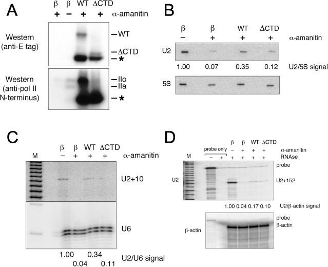
U small nuclear RNAs (snRNAs) and mRNAs are both transcribed by RNA polymerase II (Pol II), but the snRNAs have unusual TATA-less promoters and are neither spliced nor polyadenylated; instead, 3' processing is directed by a highly conserved 3' end formation signal that requires initiation from an snRNA promoter. Here we show that the C-terminal domain (CTD) of Pol II is required for efficient U2 snRNA transcription, as it is for mRNA transcription. However, CTD kinase inhibitors, such as 5,6-dichloro-1-beta-D-ribofuranosylbenzimidazole (DRB) and 1-(5-isoquinolinesulfonyl)-2-methylpiperazine (H7), that block mRNA elongation do not affect U2 transcription, although 3' processing of the U2 primary transcript is impaired. We show further that U2 transcription is preferentially inhibited by low doses of UV irradiation or actinomycin D, which induce CTD kinase activity, and that UV inhibition can be rescued by treatment with DRB or H7. We propose that Pol II complexes transcribing snRNAs and mRNAs have distinct CTD phosphorylation patterns. mRNA promoters recruit factors including kinases that hyperphosphorylate the CTD, and the CTD in turn recruits proteins needed for mRNA splicing and polyadenylation. We predict that snRNA promoters recruit factors including a CTD kinase(s) whose snRNA-specific phosphorylation pattern recruits factors required for promoter-coupled 3' end formation.


Figures

FIG. 1.
FIG. 1.
U2 snRNA constructs, primers, and probes used in the analysis. DSE, distal sequence element; PSE, proximal sequence element; 3′ box, 3′ end formation signal. Sequence elements upstream of the mature U2 snRNA coding region are indicated by negative numbers, and those downstream are indicated by positive numbers. The slanting line indicates 67 nt of vector sequence at the 5′ end of the RNase protection probe.
FIG. 2.
FIG. 2.
U2 snRNA transcription and 3′ processing require the CTD of Pol II. (A) Transient expression in HT1080 cells of E-tagged, α-amanitin-resistant Pol II LS constructs with (WT) or without (ΔCTD) an intact CTD. As assayed by Western blotting against the E-tag (upper panel), the WT construct generates an intact, phosphorylated LS (IIo); the ΔCTD construct generates comparable levels of a truncated LS; both the WT and ΔCTD constructs generate large amounts of an N-terminal LS fragment (asterisk), as seen previously (14, 24, 32); and no tagged LS is seen upon transfection with a control β-galactosidase expression construct. Assayed by Western blotting against the N terminus of Pol II (lower panel), the α-amanitin-resistant LS constructs are vastly overexpressed compared to endogenous Pol II (compare right two lanes with left two), and endogenous Pol II is degraded after exposure to 2 μg of α-amanitin/ml for 24 h (compare left two lanes). (B) Nascent U2 snRNA assayed by run-on transcription, using 5S rRNA as an internal control. Cells were harvested 24 h after transfection, and nuclear run-on transcription was conducted in the presence or absence of α-amanitin (2 μg/ml). (C) U2+10 precursor assayed by primer extension. α-Amanitin was added 24 h after transfection, and cells were incubated for an additional 24 h to degrade endogenous Pol II LS before harvesting RNA. (D) U2 snRNA primary transcript assayed by RNase protection. RNA was harvested as for panel C and assayed using the probe shown in panel A. The U2+152 signal reflects transcripts extending beyond position +152 downstream of the U2 coding region. The small amount of full-length probe seen reflects incomplete RNase digestion. RNase protection conducted with RNase A as well as T1 results in the absence of undigested probe and sharper U2+152 bands but also reveals a spurious band at U2+110 (data not shown), perhaps due to an AT-rich region in this area. Minor degradation of the high-specific-activity probe reflects autoradiolysis and does not affect our results because the probe cannot protect itself (see lane with probe only plus RNase).
FIG. 3.
FIG. 3.
Effect of CTD kinase inhibitors on U2 snRNA transcription and 3′ processing. (A) Western blots against the N terminus of the Pol II LS. CTD kinase inhibitors DRB, H7, and H8 cause a rapid shift of the Pol II LS from the hyperphosphorylated form (IIo) to the hypophosphorylated form (IIa). (B) U2 transcription assayed by nuclear run-on. U2+10 levels are not significantly affected by DRB or H7. (C) U2+10 precursor detected by primer extension, using U6 as an internal control. The U2+10 and U6 primer extensions were performed in a single reaction and resolved on a single gel; the upper panel of this gel (U2+10) was exposed for longer than the lower panel (U6 internal control) to reveal the weaker U2+10 signal. U2 snRNA is unaffected by exposure to DRB or H7 for 4 h. (D) RNase protection assay for U2 primary transcripts as performed for Fig. 1E. Significant accumulation of 3′-extended U2 species is seen upon treatment with DRB (50 μM), H7 (40 μM), or H8 (40 μM) for 4 h. As a negative control, cells were treated with a high level of ActD (2 μg/ml, 2 h) to block U2 transcription (also see Fig. 4 and 6).
FIG. 4.
FIG. 4.
Northern blot against a transiently transfected U2 maxigene. Mature U2 maxigene accumulation is reduced in the presence of the CTD kinase inhibitors DRB (50 μM) and H7 (40 μM) for 5 h. The maxigene has a 10-nt insertion within the U2 coding region; the construct has previously been shown to produce normally processed U2 (42). The oligonucleotide probe used to detect the maxigene cross-reacts weakly with endogenous mature U2, providing an internal control. As a negative control, cells were treated with a high level of ActD (2 μg/ml, 5 h) to block U2 transcription.
FIG. 5.
FIG. 5.
Effect of CTD hyperphosphorylation on U2 snRNA transcription and 3′ processing. (A) Western blot against the N terminus of the Pol II LS. A low dose of UV irradiation or ActD shifts the Pol II LS from the hypophosphorylated (IIa) form to the hyperphosphorylated (IIo) form. (B) U2 transcription, assayed by nuclear run-on, is reduced by UV irradiation (6 h) or a low dose of ActD (50 ng/ml, 2 h), whereas β-actin transcription is less severely affected. (C) U2+10 precursor, detected by primer extension, is reduced by UV or ActD treatment. U6 was used as a control as for Fig. 2C. (D) RNase protection assay for U2 primary transcripts, as in Fig. 2D. A moderate reduction in 3′-extended U2 species is seen after treatment with UV (6 h) or a low level of ActD (50 ng/ml, 2 h), whereas a high level of ActD (2 μg/ml, 2 h) completely blocks U2 transcription.
FIG. 6.
FIG. 6.
Effects of CTD kinase inhibitors on UV inhibition of U2 snRNA transcription and 3′ processing. (A) As shown by Western blotting with an antibody against the N terminus of Pol II LS, DRB (50 μM) and H7 (40 μM) shift the distribution of Pol II LS to the hypophosphorylated (IIa) form and prevent UV-mediated redistribution of Pol II LS to the hyperphosphorylated (IIo) form. (B) U2+10 precursor as detected by primer extension is drastically reduced after UV irradiation but restored in the presence of DRB (50 μM) or H7 (40 μM). (C) As shown by nuclear run-on, UV inhibition of U2 transcription 6 h postirradiation is rescued by DRB (50 μM) or H7 (40 μM). (D) RNase protection with the U2+152 probe complementary to the 3′ region of the primary transcript indicates accumulation of 3′-extended U2 species 6 h after UV irradiation in the presence of DRB or H7 compared to results with either untreated cells or UV-irradiated cells alone. A high dose of ActD (2 μg/ml, 2 h) served as a negative control to block all transcription.

References

    1. Adamczewski, J. P., M. Rossignol, J. P. Tassan, E. A. Nigg, V. Moncollin, and J. M. Egly. 1996. MAT1, cdk7 and cyclin H form a kinase complex which is UV light-sensitive upon association with TFIIH. EMBO J. 15:1877-1884. - PMC - PubMed
    1. Allison, L. A., M. Moyle, M. Shales, and C. J. Ingles. 1985. Extensive homology among the largest subunits of eukaryotic and prokaryotic RNA polymerases. Cell 42:599-610. - PubMed
    1. Baskaran, R., G. G. Chiang, T. Mysliwiec, G. D. Kruh, and J. Y. Wang. 1997. Tyrosine phosphorylation of RNA polymerase II carboxyl-terminal domain by the Abl-related gene product. J. Biol. Chem. 272:18905-18909. - PubMed
    1. Baskaran, R., M. E. Dahmus, and J. Y. Wang. 1993. Tyrosine phosphorylation of mammalian RNA polymerase II carboxyl-terminal domain. Proc. Natl. Acad. Sci. USA 90:11167-11171. - PMC - PubMed
    1. Bender, K., C. Blattner, A. Knebel, M. Iordanov, P. Herrlich, and H. J. Rahmsdorf. 1997. UV-induced signal transduction. J. Photochem. Photobiol. B 37:1-17. - PubMed

Publication types

MeSH terms

LinkOut - more resources