. 2004 Jan;24(2):899-911.
doi: 10.1128/MCB.24.2.899-911.2004.

Genomic instability and enhanced radiosensitivity in Hsp70.1- and Hsp70.3-deficient mice

Affiliations

Genomic instability and enhanced radiosensitivity in Hsp70.1- and Hsp70.3-deficient mice

Clayton R Hunt et al. Mol Cell Biol. 2004 Jan.

Abstract

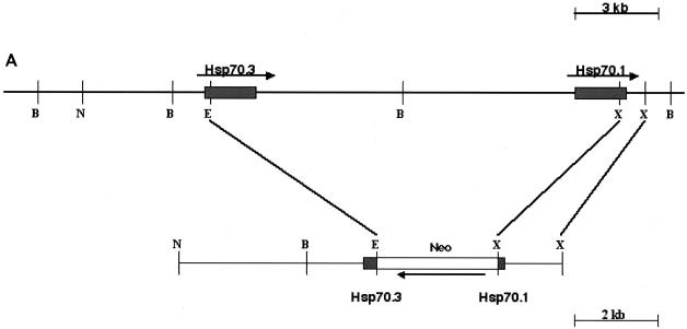
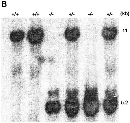
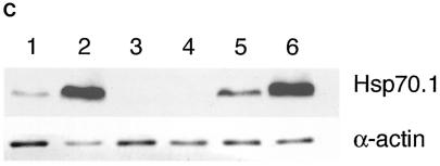
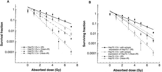
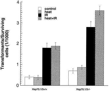
Heat shock proteins (HSPs) are highly conserved among all organisms from prokaryotes to eukaryotes. In mice, the HSP genes Hsp70.1 and Hsp70.3 are induced by both endogenous and exogenous stressors, such as heat and toxicants. In order to determine whether such proteins specifically influence genomic instability, mice deficient for Hsp70.1 and Hsp70.3 (Hsp70.1/3(-/-) mice) were generated by gene targeting. Mouse embryonic fibroblasts (MEFs) prepared from Hsp70.1/3(-/-) mice did not synthesize Hsp70.1 or Hsp70.3 after heat-induced stress. While the Hsp70.1/3(-/-) mutant mice were fertile, their cells displayed genomic instability that was enhanced by heat treatment. Cells from Hsp70.1/3(-/-) mice also display a higher frequency of chromosome end-to-end associations than do control Hsp70.1/3(+/+) cells. To determine whether observed genomic instability was related to defective chromosome repair, Hsp70.1/3(-/-) and Hsp70.1/3(+/+) fibroblasts were treated with ionizing radiation (IR) alone or heat and IR. Exposure to IR led to more residual chromosome aberrations, radioresistant DNA synthesis (a hallmark of genomic instability), increased cell killing, and enhanced IR-induced oncogenic transformation in Hsp70.1/3(-/-) cells. Heat treatment prior to IR exposure enhanced cell killing, S-phase-specific chromosome damage, and the frequency of transformants in Hsp70.1/3(-/-) cells in comparison to Hsp70.1/3(+/+) cells. Both in vivo and in vitro studies demonstrate for the first time that Hsp70.1 and Hsp70.3 have an essential role in maintaining genomic stability under stress conditions.


Figures

FIG. 1.
FIG. 1.
Targeted disruption of Hsp70.1/3. (A) Genomic structure of Hsp70.1/3 and design of targeting construct. The Hsp70 gene replacement vector was constructed by deletion of approximately 11 kb of genomic DNA separating the 5′ end of the Hsp70.3 gene and the 3′ end of the Hsp70.1 gene, thereby simultaneously inactivating both genes. This region was replaced with a 3.1-kb neo gene driven by the RNA polymerase II gene promoter. (B) Southern blot analysis of wild-type (+/+) Hsp70.1/3+/+ and correctly targeted heterozygote (+/−) Hsp70.1/3+/− and homozygote (−/−) Hsp70.1/3−/− mice, demonstrating the presence of the recombined 5.2-kb BamHI/HindIII fragment.
FIG. 1.
FIG. 1.
Targeted disruption of Hsp70.1/3. (A) Genomic structure of Hsp70.1/3 and design of targeting construct. The Hsp70 gene replacement vector was constructed by deletion of approximately 11 kb of genomic DNA separating the 5′ end of the Hsp70.3 gene and the 3′ end of the Hsp70.1 gene, thereby simultaneously inactivating both genes. This region was replaced with a 3.1-kb neo gene driven by the RNA polymerase II gene promoter. (B) Southern blot analysis of wild-type (+/+) Hsp70.1/3+/+ and correctly targeted heterozygote (+/−) Hsp70.1/3+/− and homozygote (−/−) Hsp70.1/3−/− mice, demonstrating the presence of the recombined 5.2-kb BamHI/HindIII fragment.
FIG. 2.
FIG. 2.
Northern blot analysis of Hsp70.1 and Hsp70.3 tissue-specific mRNA expression under nonstress conditions. A radiolabeled 3′ untranslated region probe derived from the mouse Hsp70.1 or Hsp70.3 gene was hybridized to fractionated poly(A) RNA isolated from eight mouse tissues. Note that Hsp70.1 and Hsp70.3 mRNA are highly expressed in kidney and lung tissue and that the major difference between Hsp70.1 and Hsp70.3 was found to be a lack of Hsp70.3 expression in liver tissue.
FIG. 3.
FIG. 3.
Synthesis of Hsp70.1 and Hsp70.3 mRNA and protein in Hsp70.1/3−/− and Hsp70.1/3+/+ MEFs. (A) Northern blot analysis of Hsp70.1 and Hsp70.3 mRNA levels. Cells were subjected to heat shock at 43°C for 30 min and then examined for mRNA expression of Hsp70.1 and Hsp70.3 with a radiolabeled DNA fragment probe derived from the mouse Hsp70.1 or Hsp70.3 gene coding region. (a) Control (Hsp70.1/3+/+) cells treatedwith heat and examined for mRNA expression. Lane 1, control RNA from unheated cells; lanes 2 to 5, cells subjected to heat shock and recovery for 1, 4, 8, and 24 h, respectively; lanes 6 to 9, cells subjected to heat shock, recovery for 24 h, heat treatment again for 30 min at 43°C, and no recovery or 1, 4, or 8 h of recovery, respectively. Note that the primary MEFs do not show detectable levels of Hsp70.1 and Hsp70.3 under nonstress conditions and that the heat shock induces both Hsp70.1 and Hsp70.3 mRNA. The induction of Hsp70.1 and Hsp70.3 is restored after a second heat shock treatment. (b) Mutant (Hsp70.1/3−/−) and control (Hsp70.1/3+/+) cells treated with heat and examined for mRNA expression. Lanes 1 to 7, Hsp70.1/3−/− cells; lane 1, control RNA from unheated cells; lanes 2 to 5, cells subjected to heat shock and no recovery or 1, 4, or 24 h of recovery, respectively; lanes 6 and 7, cells subjected to heat treatment, recovery for 24 h, heat treatment again for 30 min at 43°C, and no recovery or 2 h of recovery, respectively; lanes 8 and 9, Hsp70.1/3+/+ cells subjected to heat shock with either no or 1 h of recovery, respectively. The induction of Hsp70.1 and Hsp70.3 is restored in Hsp70.1/3+/+ but not in Hsp70.1/3−/− cells after a second heat shock treatment. (B) Heat-induced synthesis of Hsp70 in fibroblasts with and without Hsp70.1/3 genes. Polyacrylamide gel analysis of 3H-leucine-pulse-labeled (1 h) proteins synthesized after heat shock treatment. Shown is protein synthesis in Hsp70.1/3+/+ and Hsp70.1/3−/− cells (a) and Hsp70.1/3−/− cells without and with ectopically expressing Hsp70.1 (b). Cells were subjected to heat shock at 43°C for 30 min and no further treatment (lane 1); or recovery for 24 h (lane 2); or recovery for 24 h and a second heat shock at 43°C for 30 min (lane 3) or recovery for 1 h (lane 4). Note the appearance of the 70-kDa protein in lanes 2 and 4 representing cell lysates of Hsp70.1/3+/+ and Hsp70.1/3−/− cells with ectopically expressed Hsp70.1. The arrow indicates the appearance of the 70-kDa protein in Hsp70.1/3+/+ and Hsp70.1/3−/− cells with ectopically expressed Hsp70.1, while no such band is found in Hsp70.1/3−/− cells. (C) Western blot analysis for HSP70 synthesis following heat shock as detected by anti-HSP70 antibody. Cells were subjected to heat shock at 43°C for 30 min (with 2 h of recovery in lanes 2, 4, and 6) and examined for HSP70 protein with anti-HSP70 antibody. Lanes: 1 and 2, Hsp70.1/3+/+ cells; 3 and 4, Hsp70.1/3−/− cells; 5 and 6, Hsp70.1/3−/− cells with ectopically expressed Hsp70.1. Note the increase in HSP70 protein in lanes 2 and 6 whereas no HSP70 was detected in lanes 3 and 4.
FIG. 3.
FIG. 3.
Synthesis of Hsp70.1 and Hsp70.3 mRNA and protein in Hsp70.1/3−/− and Hsp70.1/3+/+ MEFs. (A) Northern blot analysis of Hsp70.1 and Hsp70.3 mRNA levels. Cells were subjected to heat shock at 43°C for 30 min and then examined for mRNA expression of Hsp70.1 and Hsp70.3 with a radiolabeled DNA fragment probe derived from the mouse Hsp70.1 or Hsp70.3 gene coding region. (a) Control (Hsp70.1/3+/+) cells treatedwith heat and examined for mRNA expression. Lane 1, control RNA from unheated cells; lanes 2 to 5, cells subjected to heat shock and recovery for 1, 4, 8, and 24 h, respectively; lanes 6 to 9, cells subjected to heat shock, recovery for 24 h, heat treatment again for 30 min at 43°C, and no recovery or 1, 4, or 8 h of recovery, respectively. Note that the primary MEFs do not show detectable levels of Hsp70.1 and Hsp70.3 under nonstress conditions and that the heat shock induces both Hsp70.1 and Hsp70.3 mRNA. The induction of Hsp70.1 and Hsp70.3 is restored after a second heat shock treatment. (b) Mutant (Hsp70.1/3−/−) and control (Hsp70.1/3+/+) cells treated with heat and examined for mRNA expression. Lanes 1 to 7, Hsp70.1/3−/− cells; lane 1, control RNA from unheated cells; lanes 2 to 5, cells subjected to heat shock and no recovery or 1, 4, or 24 h of recovery, respectively; lanes 6 and 7, cells subjected to heat treatment, recovery for 24 h, heat treatment again for 30 min at 43°C, and no recovery or 2 h of recovery, respectively; lanes 8 and 9, Hsp70.1/3+/+ cells subjected to heat shock with either no or 1 h of recovery, respectively. The induction of Hsp70.1 and Hsp70.3 is restored in Hsp70.1/3+/+ but not in Hsp70.1/3−/− cells after a second heat shock treatment. (B) Heat-induced synthesis of Hsp70 in fibroblasts with and without Hsp70.1/3 genes. Polyacrylamide gel analysis of 3H-leucine-pulse-labeled (1 h) proteins synthesized after heat shock treatment. Shown is protein synthesis in Hsp70.1/3+/+ and Hsp70.1/3−/− cells (a) and Hsp70.1/3−/− cells without and with ectopically expressing Hsp70.1 (b). Cells were subjected to heat shock at 43°C for 30 min and no further treatment (lane 1); or recovery for 24 h (lane 2); or recovery for 24 h and a second heat shock at 43°C for 30 min (lane 3) or recovery for 1 h (lane 4). Note the appearance of the 70-kDa protein in lanes 2 and 4 representing cell lysates of Hsp70.1/3+/+ and Hsp70.1/3−/− cells with ectopically expressed Hsp70.1. The arrow indicates the appearance of the 70-kDa protein in Hsp70.1/3+/+ and Hsp70.1/3−/− cells with ectopically expressed Hsp70.1, while no such band is found in Hsp70.1/3−/− cells. (C) Western blot analysis for HSP70 synthesis following heat shock as detected by anti-HSP70 antibody. Cells were subjected to heat shock at 43°C for 30 min (with 2 h of recovery in lanes 2, 4, and 6) and examined for HSP70 protein with anti-HSP70 antibody. Lanes: 1 and 2, Hsp70.1/3+/+ cells; 3 and 4, Hsp70.1/3−/− cells; 5 and 6, Hsp70.1/3−/− cells with ectopically expressed Hsp70.1. Note the increase in HSP70 protein in lanes 2 and 6 whereas no HSP70 was detected in lanes 3 and 4.
FIG. 4.
FIG. 4.
Effect of Hsp70.1/3 inactivation on cell growth. MEFs were seeded in plates, and cell counts were determined at regular intervals. Numbers of cells are plotted against days of growth in a semilog diagram. MEFs without Hsp70.1/3 (passages 3 and 7) exhibit slightly slower growth kinetics than do parental wild-type cells, and the differences in growth kinetics are significant (P < 0.05). Atm-null fibroblasts were used as a positive control to determine the growth abnormalities.
FIG. 5.
FIG. 5.
Telomerase activity and telomere signals. (A) Comparison of telomerase activities in MEFs with and without Hsp70.1/3. Note that Hsp70.1/3−/− cells have a lower telomerase activity per unit of protein than do Hsp70.1/3+/+ cells and that the differences in telomerase activity are significant (P < 0.01). Ectopic expression of Hsp70.1 restored telomerase activity in Hsp70.1/3−/− cells, almost to the level in Hsp70.1/3+/+ cells. (B) Segments of metaphases from Hsp70.1/3+/+ and Hsp70.1/3−/− cells showing telomere FISH signals. Hsp70.1/3+/+ (a) and Hsp70.1/3−/− (b and c) cells were analyzed by FISH with a telomere-specific probe. Note the telomere associations (indicated by arrows in panel b) as well as breaks near telomeres (indicated by the arrow in panel c) in Hsp70.1/3−/− cells.
FIG. 6.
FIG. 6.
Influence of Hsp70.1/3 inactivation on cell survival after IR and heat-IR treatment. (A) Dose-response curves are shown for cells with and without Hsp70.1/3. Cells were treated with IR while growing exponentially and asynchronously. Hsp70.1/3−/− cells were more sensitive to cell killing by IR than were wild-type cells, and the differences are significant (P < 0.05). Cells were also subjected to heat treatment at 43°C for 30 min and then irradiated with different doses of IR. Hsp70.1/3−/− cells are more sensitive to cell killing after heat and IR treatment than are Hsp70.1/3+/+ cells. (B) Ectopic expression of Hsp70.1 in Hsp70.1/3−/− cells rescued the enhanced killing by IR or heat-modulated IR-induced cell killing.
FIG. 7.
FIG. 7.
Chromosomal aberrations after heat, IR, and heat-IR treatment in cells with and without Hsp70.1/3. (A) Cells in plateau phase either were subjected to heat treatment at 43°C for 30 min or irradiated with 3 Gy or were first treated with heat at 43°C for 30 min followed by irradiation with 3 Gy, incubated for 18 h postirradiation, and then subcultured, and metaphases were collected. G1-type aberrations were examined at metaphase. Categories of asymmetric chromosome aberrations scored included dicentrics, centric rings, interstitial deletions-acentric rings, and terminal deletions. Treatment with heat alone did not induce G1-type chromosome aberrations. The frequency of chromosomal aberrations was higher in samples treated with heat and IR than in samples treated with IR only; however, the differences were not statistically significant. (B) Cells in exponential phase were treated with heat at 43°C for 30 min or irradiated with 2 Gy or first treated with heat at 43°C for 30 min followed by irradiation with 2 Gy. Metaphases were harvested after 3 h following irradiation and examined for chromosomal aberrations. The difference between chromatid as well as chromosomal aberrations induced by IR and those induced by heat plus IR is significantly higher in Hsp70.1/3−/− cells (P < 0.01). (C) Cells in exponential phase were treated with heat at 43°C for 30 min or irradiated with 1 Gy or first treated with heat at 43°C for 30 min followed by irradiation with 1 Gy. Metaphases were harvested after 1 h following irradiation and examined for chromosomal aberrations. The differences in chromosomal aberrations between samples treated with IR and those treated with heat-IR are not statistically significant. Note that Hsp70.1/3−/− cells have relatively more chromosomal aberrations than do parental Hsp70.1/3+/+ cells in all phases of the cell cycle;however, Hsp70.1/3−/− cells treated with heat-IR have more S-phase-specific chromosomal aberrations than do cells treated with IR only, suggesting that Hsp70.1/3 may have a specific role in S-phase-specific DNA repair. (D) DNA synthesis after IR treatment. Asynchronously growing Hsp70.1/3−/− and Hsp70.1/3+/+ cells were irradiated at the doses indicated. The rate of DNA synthesis was determined 1 h postirradiation by pulse-labeling with [3H]thymidine for 20 min. The values of unirradiated controls were set to 100% for each cell type. The mean and standard deviation of triplicate experimental points are shown.
FIG. 8.
FIG. 8.
Influence of inactivation of Hsp70.1/3 on oncogenic transformation in vitro. Control and heat-treated cells (43°C for 30 min) with and without irradiation (1 Gy) were examined for cellular transformation. Note that heat treatment had no effect on spontaneous transformation in Hsp70.1/3+/+ cells and very little effect on IR-induced transformation, whereas heat had a profound effect on spontaneous as well as IR-induced transformation in Hsp70.1/3−/− cells. The increase in the frequency of transformants in Hsp70.1/3−/− cells is significant (P < 0.025) compared to Hsp70.1/3+/+ cells.

References

    1. Barker, K., M. Khayat, N. Miller, M. Wilson, L. W. Clem, and E. Bengten. 2002. Immortal and mortal clonal lymphocyte lines from channel catfish: comparison of telomere length, telomerase activity, tumor suppressor and heat shock protein expression. Dev. Comp. Immunol. 26:45-51. - PubMed
    1. Bercovich, B., I. Stancovski, A. Mayer, N. Blumenfeld, A. Laszlo, A. L. Schwartz, and A. Ciechanover. 1997. Ubiquitin-dependent degradation of certain protein substrates in vitro requires the molecular chaperone Hsc70. J. Biol. Chem. 272:9002-9010. - PubMed
    1. Bodnar, A. G., M. Ouellette, M. Frolkis, S. E. Holt, C. P. Chiu, G. B. Morin, C. B. Harley, J. W. Shay, S. Lichtsteiner, and W. E. Wright. 1998. Extension of life-span by introduction of telomerase into normal human cells. Science 279:349-352. - PubMed
    1. Bukau, B., E. Deuerling, C. Pfund, and E. A. Craig. 2000. Getting newly synthesized proteins into shape. Cell 101:119-122. - PubMed
    1. Bukau, B., and A. L. Horwich. 1998. The Hsp70 and Hsp60 chaperone machines. Cell 92:351-366. - PubMed

Publication types

MeSH terms