. 2003 Dec 15;17(24):3029-35.
doi: 10.1101/gad.1143403.

Similar MLL-associated leukemias arising from self-renewing stem cells and short-lived myeloid progenitors

Affiliations

Similar MLL-associated leukemias arising from self-renewing stem cells and short-lived myeloid progenitors

Antonio Cozzio et al. Genes Dev. .

Abstract

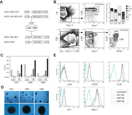
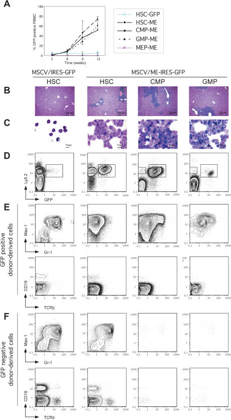
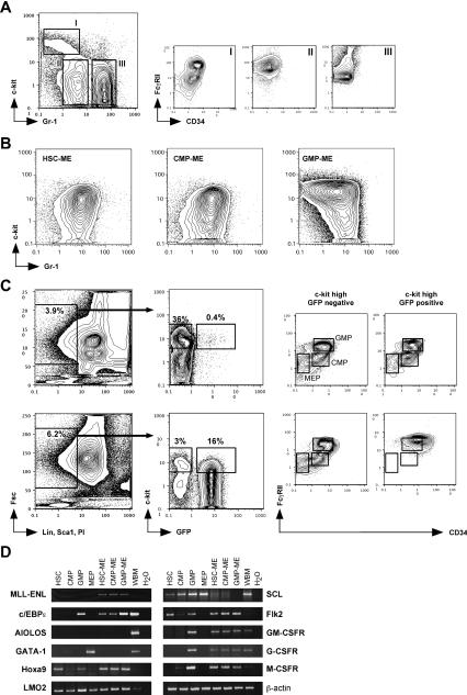
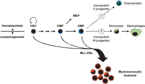
We have used the hematopoietic system as a model to investigate whether acute myeloid leukemia arises exclusively from self-renewing stem cells or also from short-lived myeloid progenitors. When transduced with a leukemogenic MLL fusion gene, prospectively isolated stem cells and myeloid progenitor populations with granulocyte/macrophage differentiation potential are efficiently immortalized in vitro and result in the rapid onset of acute myeloid leukemia with similar latencies following transplantation in vivo. Regardless of initiating cell, leukemias displayed immunophenotypes and gene expression profiles characteristic of maturation arrest at an identical late stage of myelomonocytic differentiation, putatively a monopotent monocytic progenitor stage. Our findings unequivocally establish the ability of transient repopulating progenitors to initiate myeloid leukemias in response to an MLL oncogene, and support the existence of cancer stem cells that do not necessarily overlap with multipotent stem cells of the tissue of origin.


Figures

Figure 1.
Figure 1.
Enhanced in vitro self-renewal following transduction of purified myeloid progenitors with MLL-ENL. (A) Schematic representation of the retroviral control and oncogenic MLL-ENL constructs used for the in vitro and in vivo transduction experiments. (B) Example of FACS sorting conditions used to obtain highly purified populations of HSC (lin-/Thy1.1low/c-Kithigh/Sca-1high), CMP (lin-/Thy1.1-/Il7R-/c-Kithigh/Sca-1-/CD34+/FcγRlow), GMP (lin-/Thy1.1-/Il7R-/c-Kithigh/Sca-1-/CD34high/FcγRhigh), and MEP (lin-/Thy1.1-/Il7R-/c-Kithigh/Sca-1-/CD34-/FcγRlow). In methylcellulose CFU assay, both HSC and CMP gave rise to the entire range of myeloid and erythroid colonies including mix colonies (Mix), whereas GMP only gave rise to colonies composed of macrophages (M) and/or granulocytes (GM and G) and MEP to colonies composed of erythrocytes (E) and/or megakaryocytes (E/Meg and Meg), thereby attesting the purity of the starting populations. (C) In vitro immortalization of HSC and committed CMP and GMP progenitors. Bar graph indicates the numbers of colonies obtained after each round of plating in methylcellulose supplemented with IL-3, IL-6, SCF, GM-CSF, and G-418 for the first round only, and are representative of three independent experiments. Transduction efficiencies can be calculated from the first round of plating as the number of drug-resistant positively selected colonies divided by the numbers of starting cells (HSC: vector 30.3%, ME 22.7%; CMP: vector 80%, ME 26.3%; GMP: vector 77.1%, ME 50%). (D) Typical colonies showing blast morphology obtained after MLL-ENL-transduction of HSC, CMP, and GMP populations (upper panel: 40× magnification; lower panel: 200× magnification). (E) Immunophenotype characterization of the ME-transduced HSC, CMP, and GMP cell lines showing similar expression levels of the c-Kit, CD34, FcγR, Gr-1, and Mac-1 surface markers.
Figure 2.
Figure 2.
Acute myeloid leukemias induced by MLL-ENL from stem and progenitor cells. (A) The extent of engraftment and development of AML was monitored by FACS analysis of GFP+ cells in peripheral blood mononuclear cells (PBMC). Mice (five per cohort) were transplanted with 1000 HSC transduced with control (blue line) or ME (dashed line) retroviruses, or with 10,000 ME-transduced CMP (black line), GMP (dotted line), or MEP (red line). Mice transplanted with control-transduced CMP, GMP, or MEP did not show any donor-derived Ly5.2+ cells after 6 to 7 wk posttransplantation (data not shown). (B-C) AML in preterminally leukemic mice. (B) Liver histology showing massive blast invasion in recipients of ME-transduced stem and progenitor cells. (C) May-Grünwald-Giemsa staining of splenocytes, indicating mostly blast and myelomonocytic morphologies. (D-E) Chimera analysis in mice 9 wk after transplantation of transduced stem and progenitor populations. ACK-treated bone marrow cells were analyzed for expression of GFP, Ly5.2 (donor-derived), and various myeloid (Mac-1, Gr-1) and lymphoid (CD19, TCRβ) markers. (D) An expanded population of ME-transduced donor-derived GFP+ cells is present in all leukemic mice. (E) ME-transduced GFP+ donor-derived cells only expressed myeloid markers (Mac-1high, Gr-1-/low) in contrast to multilineage readout of control-transduced HSC. (F) Multilineage reconstitution from untransduced GFP- donor-derived cells in ME-transduced HSC reconstituted mice. Consistent with their transient repopulating ability, untransduced GFP- donor-derived cells are not detected in CMP or GMP transplanted mice, confirming the absence of HSC contamination in the transplant.
Figure 3.
Figure 3.
Characterization of the leukemic cells. (A) Normal bone marrow stained with c-Kit and Gr-1 shows three distinct populations: a c-Kitint./high/Gr-1-/low population (I), which is CD34+/FcγR+ and contains the myeloid CMP and GMP progenitors (Akashi et al. 2000); a c-Kit-/Gr-1low population (II), which is CD34-/FcγRhigh and contains the myelomonocytic cells; and a c-Kit-/Gr-1high population (III), which is CD34-/FcγRint. and contains the neutrophilic granulocytes (Lagasse and Weissman 1996). (B) In leukemic mice, all ME-transduced GFP+ donor-derived cells from HSC, CMP, and GMP origin do not show the immunophenotypes of the starting populations, but are consistently c-Kitlow-int./Gr-1int.. (C) Mice transplanted with control-(upper panel) or ME-(lower panel) transduced HSC were analyzed for GFP expression in myeloid progenitor compartments at 9 wk posttransplantation. In control-transduced mice, c-Kithigh/GFP+ donor-derived cells contributed to all myeloid progenitor populations, whereas in leukemic mice the same population only yielded leukemic cells with a FcγRIIhigh/CD34+ phenotype. (D) Gene expression analyses comparing HSC and myeloid progenitors to their ME-transduced leukemic counterparts. RT-PCR was performed on 100 sorted cells or 5 × 10⁵ total cells for control whole bone marrow (WBM).
Figure 4.
Figure 4.
Proposed model for MLL-ENL initiated leukemogenesis. In normal hematopoiesis, only HSC possess unlimited self-renewal potential. Differentiation occurs through a phenotypically and functionally defined series of progenitor populations with restricted half-lives and lineage potentials. Myelomonocytic leukemias induced by MLL-ENL can arise in both self-renewing HSC and short-lived progenitor cells with GM differentiation potential. Introduction of MLL-ENL uniformly leads to the accumulation of leukemic cells with the phenotype of cells immediately downstream of GMP, putatively a monopotent monocytic progenitor stage.

References

    1. Akashi K., Traver, D., Miyamoto, T., and Weissman, I.L. 2000. A clonogenic common myeloid progenitor that gives rise to all myeloid lineages. Nature 404: 193-197. - PubMed
    1. Armstrong S.A., Staunton, J.E., Silverman, L.B., Pieters, R., den Boer, M.L., Minden, M.D., Sallan, S.E., Lander, E.S., Golub, T.R., and Korsmeyer, S.J. 2002. MLL translocations specify a distinct gene expression profile that distinguishes a unique leukemia. Nat. Genet. 30: 41-47. - PubMed
    1. Armstrong S.A., Kung, A.L., Mabon, M.E., Silverman, L.B., Stam, R.W., Den Boer, M.L., Pieters, R., Kersey, J.H., Sallan, S.E., Fletcher, J.A., et al. 2003. Inhibition of FLT3 in MLL. Validation of a therapeutic target identified by gene expression based classification. Cancer Cell 3: 173-183. - PubMed
    1. Ayton P.M. and Cleary, M.L. 2001. Molecular mechanisms of leukemogenesis mediated by MLL fusion proteins. Oncogene 20: 5695-5707. - PubMed
    1. ____. 2003. Transformation of myeloid progenitors by MLL oncoproteins is dependent on Hoxa7 and Hoxa9. Genes & Dev. 17: 2298-2307. - PMC - PubMed

Publication types

MeSH terms

Substances