

Enhanced hippocampal noradrenaline and serotonin release in galanin-overexpressing mice after repeated forced swimming test
- PMID: 14701907
- PMCID: PMC314189
- DOI: 10.1073/pnas.0307042101
Enhanced hippocampal noradrenaline and serotonin release in galanin-overexpressing mice after repeated forced swimming test
Abstract
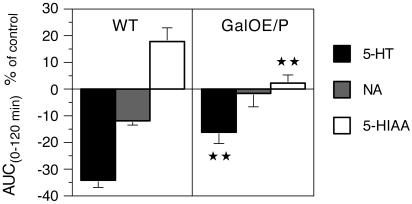
Basal and forced swimming (FS) stress-induced release of noradrenaline (NA) and serotonin (5-HT) were determined by in vivo microdialysis in the ventral hippocampus of mice overexpressing galanin under the platelet-derived growth factor B promoter (GalOE/P) or under the dopamine beta-hydroxylase promoter (GalOE/D) (only NA). WT mice served as controls. Intraventricular infusion of galanin significantly reduced basal extracellular NA in WT mice and in GalOE/P mice (albeit less so). Microdialysis sampling during a 10-min FS showed that NA and 5-HT release were elevated to 213% and 156%, respectively, in the GalOE/P group, whereas in the WT group the increases were only 127% and 119%, respectively. The second (repeated) 10-min FS (RFS) caused a marked enhancement of NA and 5-HT release in the GalOE/P mice to 344% and 275%, respectively. However, the RFS caused only a 192% increase of extracellular NA levels in the GalOE/D mice. Pretreatment with the putative peptidergic galanin receptor antagonist M35 almost completely blocked the elevation of NA and 5-HT levels in the GalOE/P after RFS. These results suggest that the NA and 5-HT hippocampal afferents in GalOE/P mice are hypersensitive to both conditioned and unconditioned stressful stimuli, such as FS, and that this effect is mediated by galanin receptors. The present findings support a role of galanin in the regulation of release of NA and 5-HT, two neurotransmitters involved in mood control.
Figures
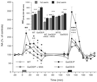
 , P < 0.05; , P < 0.01; , P < 0.002, n = 5). The galanin antagonist M35 given i.c.v. to GalOE/P mice significantly attenuated the elevation of NA levels induced by both FS and RFS stressors (
 , P < 0.05; , P < 0.001, n = 4–5). After FS, the AUC for NA in the GalOE/P mice was significantly higher than in the WT and GalOE/D mice (
 , P < 0.01, n = 5), whereas M35 completely abolished the stress effects induced by FS (, P < 0.001) and RFS (⋆⋆⋆, P < 0.001) in GalOE/P mice (Inset). M35 given to WT mice significantly reduced the AUC for NA (
 , P < 0.001; , P < 0.05; n = 5). The RFS caused a significant increase in AUC of NA in GalOE/P as compared to FS in the same group (⋆, P < 0.05) and compared to the AUC values after RFS in the WT and GalOE/D mice, respectively (⋆⋆⋆, P < 0.001).
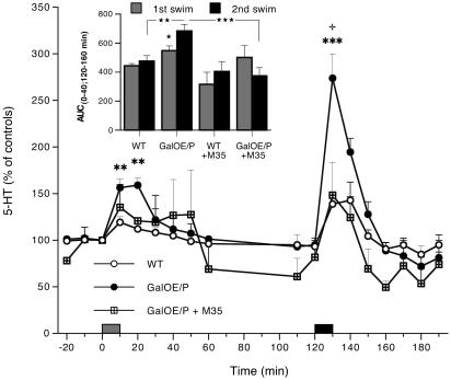
References
-
- Tatemoto, K., Rökaeus, Å., Jörnvall, H., McDonald, T. J. & Mutt, V. (1983) FEBS Lett. 164, 124-128. - PubMed
-
- Skofitsch, G. & Jacobowitz, D. M. (1985) Peptides 6, 509-546. - PubMed
-
- Melander, T., Hökfelt, T. & Rökaeus, Å. (1986) J. Comp. Neurol. 248, 475-517. - PubMed
-
- Perez, S. E., Wynick, D., Steiner, R. A. & Mufson, E. J. (2001) J. Comp. Neurol. 434, 158-185. - PubMed
Publication types
MeSH terms
Substances
LinkOut - more resources
Full Text Sources
Molecular Biology Databases
