Comparative Study
. 2004 Jan 13;101(2):452-7.
doi: 10.1073/pnas.2533829100. Epub 2003 Dec 30.

Bacillus subtilis RecU protein cleaves Holliday junctions and anneals single-stranded DNA

Affiliations
Comparative Study

Bacillus subtilis RecU protein cleaves Holliday junctions and anneals single-stranded DNA

Silvia Ayora et al. Proc Natl Acad Sci U S A. .

Erratum in

  • Proc Natl Acad Sci U S A. 2009 Jan 13;106(2):664.. Doncel, Ernesto [corrected to Doncel-Perez, Ernesto]

Abstract

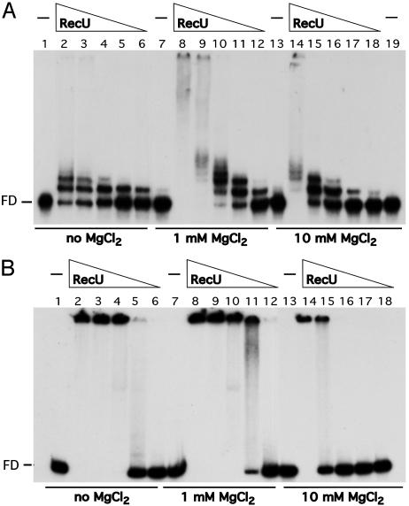
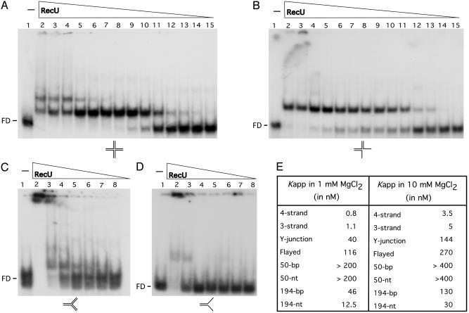
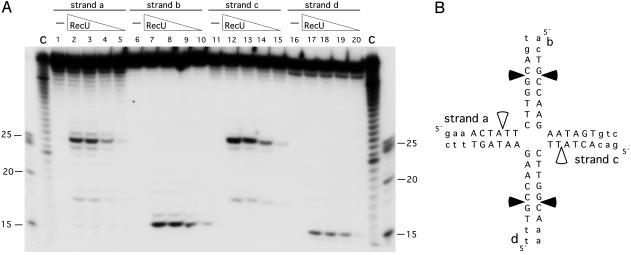
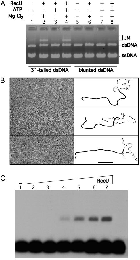
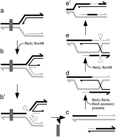
Bacillus subtilis RecU protein is involved in homologous recombination, DNA repair, and chromosome segregation. Purified RecU binds preferentially to three- and four-strand junctions when compared to single-stranded DNA (ssDNA) and double-stranded DNA (dsDNA) ( approximately 10- and approximately 40-fold lower efficiency, respectively). RecU cleaves mobile four-way junctions but fails to cleave a linear dsDNA with a putative cognate site, a finding consistent with a similar genetic defect observed for genes classified within the epsilon epistatic group (namely ruvA, recD, and recU). In the presence of Mg(2+), RecU also anneals a circular ssDNA and a homologous linear dsDNA with a ssDNA tail and a linear ssDNA and a homologous supercoiled dsDNA substrate. These results suggest that RecU, which cleaves recombination intermediates with high specificity, might also help in their assembly.


Figures

Fig. 1.
Fig. 1.
RecU binds ssDNA and dsDNA. A 194-nt γ-32P ssDNA (A) or 194-bp α-32P dsDNA (B) (0.3 μM) was incubated with various amounts of RecU (150 to 9 nM in lanes 2–6, 8–12, and 14–18 of A, and 300 to 18 nM in lanes 6–2, 8–12, and 14–18 of B) in buffer B containing no MgCl2 or the indicated amounts of MgCl2 at 37°C for 15 min. The protein·DNA complexes formed were analyzed by using EMSA. FD, free DNA; –, absence of RecU.
Fig. 2.
Fig. 2.
DNA-binding specificity of RecU. EMSA showing binding of RecU to the indicated γ-32P DNA substrates (four- and three-way, Y-junction, and flayed; AD, respectively). Binding reactions contained 0.3 μM DNA species and increasing amounts of RecU protein (from 400 to 0.05 in lanes 2–15 of A and B, and 400 to 6.2 in lanes 2–8 of C and D) in buffer B containing 1 mM MgCl2.(E) The RecU concentration to reach half-saturation (Kapp) with the different DNA substrates used at the MgCl2 concentrations indicated. The values are the average of three independent experiments.
Fig. 3.
Fig. 3.
Determination of the cleavage sites produced by RecU. (A) Four-strand junction Jbm6, γ-32P-labeled at the indicated strand, was incubated with increasing amounts of RecU (100 to 12.5 nM in lanes 2–5, 7–10, 12–15, and 17–20) in buffer B containing 10 mM MgCl2 at 37°C for 30 min. Reaction products were analyzed by using 15% denaturing PAGE. The γ-32P-labeled markers are degraded 40 (C), 25, 20 and 15 nt. (B) The sequence of the 13-bp mobile core is shown in capital letters, and the nonmobile part of the junction is shown in lowercase letters. Major nicking sites are denoted by filled arrows, and minor nicking sites are denoted by open arrows.
Fig. 4.
Fig. 4.
Formation of JMs promoted by RecU. The strand exchange reaction mixtures contained RecU (250 nM), circular M13mp18 ssDNA (30 μM), and M13mp18 linear dsDNA (60 μM) in buffer B in the presence or absence of 1 mM MgCl2 or 2 mM ATP (A). The linear 3′-tailed (A, lanes 1–4, and B) and blunt-ended dsDNA substrate (A, lanes 5–8) were used. The reaction mixture was incubated at 30°C for 30 min, and the deproteinized products were analyzed by using 0.8% AGE (A). + and –, Presence of absence of the indicated product. (B) RecU (250 nM), circular M13mp18 ssDNA (28 μM), and M13mp18 linear 3′-tailed dsDNA (28 μM) in buffer B in the presence of 1 mM MgCl2. The reaction mixture was incubated at 30°C for 30 min, and the deproteinized products were analyzed by using electron microscopy. A gallery of partially dsDNA circles with a dsDNA and ssDNA branch attached to the circle is indicated. A schematic representation of the branch of dsDNA and ssDNA is denoted. (Scale bar, 0.5 μm.) (C) A homologous 194-nt γ-32P ssDNA (0.4 μM) was incubated with supercoiled RF M13mp18 DNA (30 μM) and various concentrations of RecU (4–125 nM in lanes 2–7) in buffer B containing 1 mM MgCl2 at 37°C for 30 min. The deproteinized products were analyzed by using 0.8% AGE.
Fig. 5.
Fig. 5.
Processing of a HJ at a stalled replication fork by RuvAB and RecU. The fork was halted at a lesion (filled rectangle) within the template DNA (a) (–6). RuvAB and RecU form a four-stranded HJ (b). RecU introduces symmetrically related nicks to release a free duplex DNA end (b′). The lesion is removed (c). A free DNA end can be recombined with a homologous duplex to create a D-loop (d). RuvAB targeted to the junction site promotes branch migration and RecU resolves the junction (e and e′) (–6). The RecU cleavage in two of the four strands of the HJ restores a competent replication fork (e′). Half arrows on the DNA strands denote 3′ ends.

Similar articles

Cited by

References

    1. Kowalczykowski, S. C. (2000) Trends Biochem. Sci. 25, 156–165. - PubMed
    1. Michel, B. (2000) Trends Biochem. Sci. 25, 173–178. - PubMed
    1. Cox, M. M., Goodman, M. F., Kreuzer, K. N., Sherratt, D. J., Sandler, S. J. & Marians, K. J. (2000) Nature 404, 37–41. - PubMed
    1. McGlynn, P. & Lloyd, R. G. (2002) Nat. Rev. Mol. Cell Biol. 3, 859–870. - PubMed
    1. McGlynn, P. & Lloyd, R. G. (2002) Trends Genet. 18, 413–419. - PubMed

Publication types

LinkOut - more resources