. 2004 Jan;134(1):320-31.
doi: 10.1104/pp.103.027888. Epub 2003 Dec 30.

Pseudomonas aeruginosa-plant root interactions. Pathogenicity, biofilm formation, and root exudation

Affiliations

Pseudomonas aeruginosa-plant root interactions. Pathogenicity, biofilm formation, and root exudation

Travis S Walker et al. Plant Physiol. 2004 Jan.

Abstract

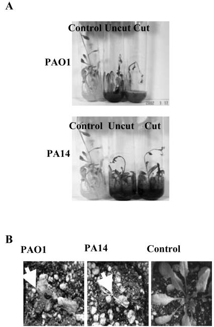
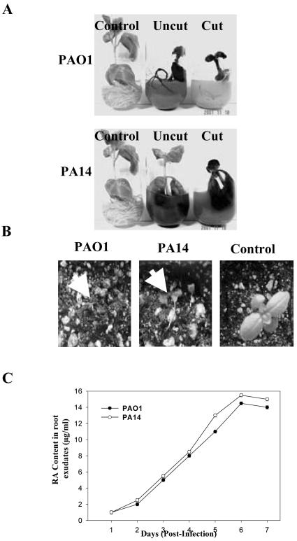
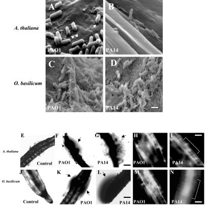
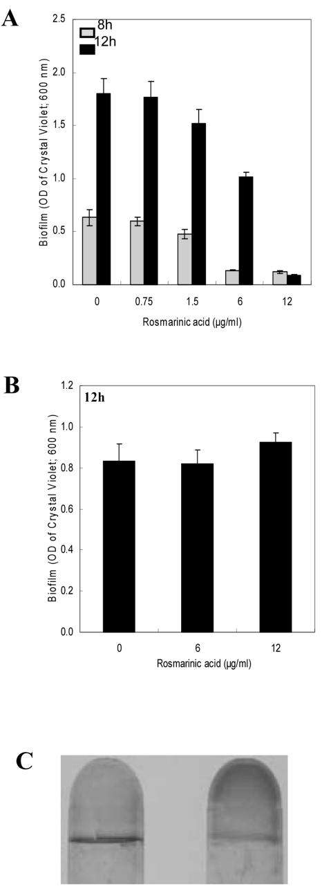
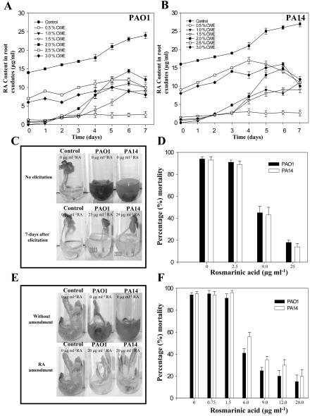
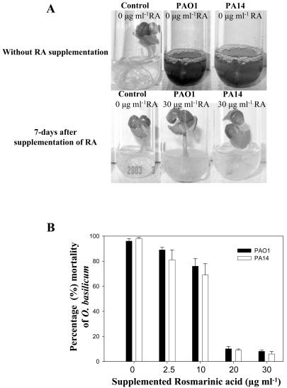
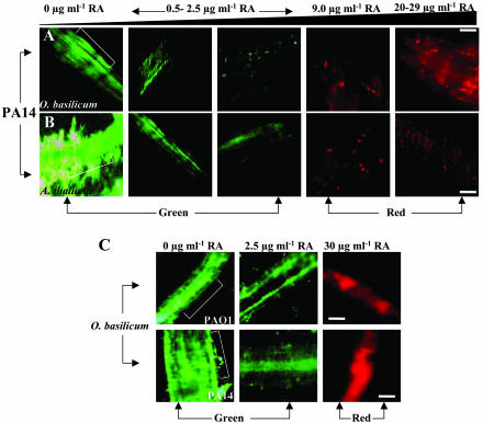
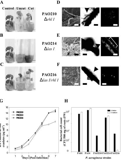
Pseudomonas aeruginosa is an opportunistic human pathogen capable of forming a biofilm under physiological conditions that contributes to its persistence despite long-term treatment with antibiotics. Here, we report that pathogenic P. aeruginosa strains PAO1 and PA14 are capable of infecting the roots of Arabidopsis and sweet basil (Ocimum basilicum), in vitro and in the soil, and are capable of causing plant mortality 7 d postinoculation. Before plant mortality, PAO1 and PA14 colonize the roots of Arabidopsis and sweet basil and form a biofilm as observed by scanning electron microscopy, phase contrast microscopy, and confocal scanning laser microscopy. Upon P. aeruginosa infection, sweet basil roots secrete rosmarinic acid (RA), a multifunctional caffeic acid ester that exhibits in vitro antibacterial activity against planktonic cells of both P. aeruginosa strains with a minimum inhibitory concentration of 3 microg mL(-1). However, in our studies RA did not attain minimum inhibitory concentration levels in sweet basil's root exudates before P. aeruginosa formed a biofilm that resisted the microbicidal effects of RA and ultimately caused plant mortality. We further demonstrated that P. aeruginosa biofilms were resistant to RA treatment under in vivo and in vitro conditions. In contrast, induction of RA secretion by sweet basil roots and exogenous supplementation of Arabidopsis root exudates with RA before infection conferred resistance to P. aeruginosa. Under the latter conditions, confocal scanning laser microscopy revealed large clusters of dead P. aeruginosa on the root surface of Arabidopsis and sweet basil, and biofilm formation was not observed. Studies with quorum-sensing mutants PAO210 (DeltarhlI), PAO214 (DeltalasI), and PAO216 (DeltalasI DeltarhlI) demonstrated that all of the strains were pathogenic to Arabidopsis, which does not naturally secrete RA as a root exudate. However, PAO214 was the only pathogenic strain toward sweet basil, and PAO214 biofilm appeared comparable with biofilms formed by wild-type strains of P. aeruginosa. Our results collectively suggest that upon root colonization, P. aeruginosa forms a biofilm that confers resistance against root-secreted antibiotics.


Figures

Figure 1.
Figure 1.
. Virulence of P. aeruginosa strains PAO1 and PA14 against Arabidopsis under in vitro and soil conditions. A, PAO1 and PA14 were infiltrated into the liquid media of cut and uncut plants, and disease symptoms and plant mortality were recorded after 7 d. B, Bacteria were also added to sterile soil of uncut plants and disease symptoms were again recorded after 7 d (arrows indicate aerial tissue damage leading to plant mortality).
Figure 2.
Figure 2.
Virulence of P. aeruginosa strains PAO1 and PA14 against sweet basil under in vitro and soil conditions. A, PAO1 and PA14 were infiltrated into the liquid media of cut and uncut plants, and disease symptoms and plant mortality were recorded after 7 d. B, Bacteria were also added to sterile soil of uncut plants and disease symptoms were again recorded after 7 d (arrows indicate aerial tissue damage leading to plant mortality). C, Increased levels of RA (micrograms per milliliter) in the root exudates of sweet basil plants infected with P. aeruginosa was determined daily by HPLC analysis upon infection with PAO1 and PA14.
Figure 3.
Figure 3.
SEM images of Arabidopsis roots infected with strains PAO1 (A) and PA14 (B) attaching perpendicularly to the root cell wall and forming a biofilm layer (arrows in A depict a perforation made by the bacterial strains). SEM of sweet basil roots with PAO1 (C) and PA14 (D) forming a mature biofilm 4 d postinoculation. Scale bar = 5 μm. Phase contrast and confocal images showing uninfected Arabidopsis roots (E) and roots infected with strains PAO1 (F and H) and PA14 (G and I); and uninfected sweet basil roots (J) and roots infected with strains PAO1 (K and M) and PA14 (L and N). Arrows indicate phase-bright material suggestive of a biofilm surrounding the roots; bracket indicates the root. Scale bar = 50 μm.
Figure 4.
Figure 4.
Effect of varying concentrations of RA on initiation of biofilm formation by strain PA14 (A) and disruption of preformed PA14 biofilms (B). A, RA was added to the (Bushnell-Haas mineral salts medium supplemented with 0.2% [w/v] dextrose and 0.5% [w/v] tryptone [BDT medium]) from the onset of the incubation period. The range of RA assessed was above and below the MIC (0, 0.75, 1.5, 6.0, and 12 μg mL-1). Biofilm formation was quantified after 8 and 12 h of incubation. B, RA was added to preformed PA14 biofilms by replacing the BDT medium with fresh medium containing RA after 8 h of incubation. The concentrations of RA tested were 0, 6.0, and 12 μgmL-1. The biofilm was then allowed to develop for an additional 4 h (to 12 h) before quantification. Error bars represent ± sd, n = 3. C, Biofilm formed in absence (left) or presence of 12 μg mL-1 of RA (right) when RA was added to the growth medium 8 h after initiation of biofilm development.
Figure 5.
Figure 5.
A and B, Influence of P. cinnamoni CWE on RA exudation in in vitro-grown cultures of sweet basil. Plants were elicited with varying concentrations CWE for 7 d before inoculation with PAO1 and PA14 (see “Materials and Methods”; values are mean ± sd, n = 5). Day 0 of inoculation corresponds to d 7 postelicitation with CWEs. C, Reduced virulence of P. aeruginosa strains PAO1 and PA14 in sweet basil plants elicited with CWE. D, Mortality rates of elicited sweet basil plants inoculated with strains PAO1 and PA14. Values are mean ± sd, n = 5. E, Reduced virulence of strains PAO1 and PA14 in RA-supplemented Arabidopsis plants compared with an untreated control. F, Mortality rates of RA-supplemented (sub-MIC and above-MIC levels) Arabidopsis plants inoculated with strains PAO1 and PA14. Values are mean ± sd, n = 5.
Figure 6.
Figure 6.
Effect of exogenous supplementation of RA to sweet basil root exudates. A, RA was added to sweet basil root exudates before infection with PAO1 and PA14 at varying concentrations (2.5-30 μg mL-1). Control plants were not supplemented with RA. Strains PAO1 and PA14 were less virulent upon supplementation with RA (30 μg mL-1). B, Mortality rates of sweet basil plants supplemented with RA (sub-MIC and above-MIC levels; 2.5-30 μg mL-1) and inoculated with strains PAO1 and PA14. B, Sweet basil plants were supplemented with RA at concentrations 10-fold greater than MIC levels. Values are mean ± sd, n = 5.
Figure 7.
Figure 7.
CSLM images of P. aeruginosa biofilms on in vitro-grown sweet basil (A) and Arabidopsis (B) roots. Bacterial viability was visualized by staining with a LIVE/DEAD BacLight Bacterial Viability kit (Molecular Probes, Eugene, OR): red areas indicate dead bacteria, and green areas indicate live bacteria (brackets represent root; scale bars = 50 μm. A, Influence of P. cinnamoni CWEs on sweet basil RA exudation and subsequent inoculation with PA14. Increasing concentrations of P. cinnamoni CWEs (2.0%-2.5%, v/v) resulted in PA14 mortality as shown in red. B, Effect of exogenous RA (0.75-20.0 μg mL-1) on PA14 biofilm formation on infected Arabidopsis roots. C, CSLM image of P. aeruginosa biofilm on in vitro-grown sweet basil roots supplemented with RA. C, Effect of exogenous RA (2.5 and 30 μg mL-1) on PAO1 and PA14 biofilm formation on infected sweet basil roots. Arabidopsis and sweet basil roots exhibited no autofluorescence.
Figure 8.
Figure 8.
Infection and mortality of sweet basil inoculated with three P. aeruginosa quorum-sensing mutants. A through C, Effect of P. aeruginosa quorum-sensing mutants in sweet basil infections assessed 7 d postinoculation. D through F, In vitro root pathogenicity in sweet basil: corresponding SEM (scale bar = 5 μm), phase contrast (scale bar = 50 μm), and CSLM images (scale bar = 50 μm). E, Strain PAO214 on sweet basil roots reveals a nearly complete biofilm compared with PAO210 and PAO216 (arrows indicate phase-bright material suggestive of a biofilm surrounding the roots; bracket indicates the root). G, RA content (micrograms per milliliter) in sweet basil root exudates infected with P. aeruginosa quorum-sensing mutants as determined daily by HPLC analysis. H, Bacterial cell counts on Arabidopsis and sweet basil roots 4 d postinfection with strains PAO1, PA14, and three quorum-sensing mutants. Average values were plotted (mean ± sd; n = 5) after inoculation with 105 bacteria per seedling. Five plants of each species were used per treatment.

References

    1. Bais HP, Loyola Vargas VM, Flores HE, Vivanco JM (2001) Root-specific metabolism: the biology and biochemistry of underground organs. In Vitro Cell Dev Biol Plant 37: 730-741
    1. Bais HP, Walker TS, Schweizer HP, Vivanco JM (2002) Root-specific elicitation and antimicrobial activity of rosmarinic acid in hairy root cultures of Ocimum basilicum. Plant Physiol Biochem 40: 983-995
    1. Chuanchuen R, Beinlich K, Hoang TT, Becher A, Karkoff-Schweizer RR, Schweizer HP (2001) Cross-resistance between triclosan and antibiotics in Pseudomonas aeruginosa is mediated by multidrug efflux pumps: exposure of a susceptible mutant strain to triclosan selects nfxB mutants over expressing MexCD-OprJ. Antimicrob Agents Chemother 45: 428-432 - PMC - PubMed
    1. Coleman FT, Mueschenborn S, Meluleni G, Ray C, Carey VJ, Vargas SO, Cannon CL, Ausubel FM, Pier GB (2003) Hypersusceptibility of cystic fibrosis mice to chronic Pseudomonas aeruginosa oropharyngeal colonization and lung infection. Proc Natl Acad Sci USA 100: 1949-1954 - PMC - PubMed
    1. Costerton JW, Lewandowski Z, Caldwell DE, Korber DR, Lappin-Scott HM (1995) Microbial biofilms. Annu Rev Microbiol 49: 711-745 - PubMed

Publication types