. 2004 Jan;36(1):100-5.
doi: 10.1038/ng1283. Epub 2003 Dec 21.

Meiotic pairing and imprinted X chromatin assembly in Caenorhabditis elegans

Affiliations

Meiotic pairing and imprinted X chromatin assembly in Caenorhabditis elegans

Christopher J Bean et al. Nat Genet. 2004 Jan.

Abstract

The genetic imprinting of individual loci or whole chromosomes, as in imprinted X-chromosome inactivation in mammals, is established and reset during gametogenesis; defects in this process in the parent can result in disease in the offspring. We describe a sperm-specific chromatin-based imprinting of the X chromosome in the nematode Caenorhabditis elegans that is restricted to histone H3 modifications. The epigenetic imprint is established during spermatogenesis and its stability in the offspring is affected by the presence of a pairing partner during meiosis in the parental germ line. We observed that DNA lacking a pairing partner during meiosis, the normal situation for the X chromosome in males, is targeted for methylation of histone H3 at Lys9 (H3-Lys9) and can be silenced. Targeting unpaired DNA for silencing during meiosis, a potential hallmark of genome defense, could therefore have a conserved role in imprinted X-chromosome inactivation and, ultimately, in sex chromosome evolution.


Conflict of interest statement

COMPETING INTERESTS STATEMENT

The authors declare that they have no competing financial interests.

Figures

Figure 1
Figure 1
The Xp chromosome lacks a subset of histone H3 modifications in the early embryo. (a,b) Antibodies to methylated H3-Lys4 (a, green) and to acetylated H3 (b, green) are excluded from one chromosome (arrowheads) in one-cell embryos counterstained with DAPI (red). Telomeric regions also appear to exclude these antibodies (small arrows). (c) Exclusion of antibody to dimethylated H3-Lys4 from a large region of the sperm pronucleus shortly after fertilization (arrowhead). (d,e) A single chromosome with reduced staining for antibody to methylated H3-Lys4 persists in two-cell (d) and four-cell (e) embryos (arrowheads). An extruded polar body (marked with an asterisk), visible in e, also stains with this antibody. (fh) Males with an X-integrated GFP transgene were mated to normal XX worms to unambiguously identify the sperm-derived X chromosome (Xp) in cross-progeny XX embryos. In representative nuclei from two-cell (f), four-cell (g) and six-cell (h) XX cross-progeny embryos, staining with antibody to dimethylated H3-Lys4 (green) is always absent from the Xp chromosome (arrowheads), marked by DNA FISH specifically targeting the transgenic sequences (blue). The FISH probe labeled approximately 50% of cross-progeny embryos, identifying XX embryos carrying both maternal and paternal X chromosomes. Red shows DAPI counterstain. Scale bars, 5 μm.
Figure 2
Figure 2
Meiotic pairing in the parent affects the stability of the epigenetic imprint in the offspring. The presence of the epigenetically imprinted X chromosome at different stages (number of nuclei) was compared in offspring from normal XO male crosses (N2 (XO)), tra-2 (q276) XX male crosses (tra-2 (XX)) and N2 hermaphrodite self-progeny (N2 (XX)). The number of embryos optically sectioned and assessed for each class and stage is indicated in parentheses at the top of each bar.
Figure 3
Figure 3
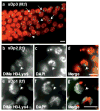
Meiotic targeting for methylation of H3-Lys9 and transcriptional repression of an unpaired X chromosome. (ad) Staining with antibody to methylated H3-Lys9 (green) in adult gonads counterstained with DAPI (red in all panels). (a) Wild-type N2 (XX) late pachytene–diplotene oocytes. Arrow indicates an individual nucleus with normal enrichment for methylated H3-Lys9 at diplotene stage in oogenesis. (b) N2 (XO) mid-pachytene spermatocytes; arrows indicate X chromosome stained with antibody to methylated H3-Lys9 present in all nuclei. (c) tra-2 (q276) XX late pachytene–diplotene spermatocytes have a pattern of staining with antibody to methylated H3-Lys9 similar to that of XX oocytes (a). Arrows indicate unstained chromosome in representative nuclei, identified by FISH costaining as the paired X homologs in the inset (arrow; blue shows DNA FISH). (d) her-1 (hv1y101) XO early- to mid-pachytene oocytes, with arrows indicating unpaired X chromosomes in several nuclei. (ej) Staining with antibody to methylated H3-Lys4 (green) in N2 (XX) germ cells initially does not label the X chromosome in pachytene cells (arrow in e) but labels all six chromosome pairs in early (f) and late (g) diakinetic oocytes. (h–j) In her-1 XO oocytes, accumulation of methylated H3-Lys4 on the unpaired X chromosome (arrows) is markedly delayed in early (i) and understained in late (j) oocytes. (kn) Strong mRNA in situ hybridization of a probe for an X-linked oocyte-enriched gene (F52D2.2; ref. 14) is detected in developing oocytes (arrows) of XX her-1 (hv1y101) worms (k), but little or no signal is observed in developing oocytes of XO worms (l). Other X-linked oocyte-enriched genes showed similar defects in transcriptional activity (data not shown). In contrast, mRNA of an autosome-linked oocyte-specific gene (emo-1) is easily detected in developing oocytes (arrows) of both XX (m) and XO (n) worms. Scale bars, 5 μm.
Figure 4
Figure 4
Targeting of methylated H3-Lys9 to the X chromosome and autosomes is determined by pairing status and not ploidy. (a,b) him-8 XX hermaphrodite pachytene-stage germ cells stained for methylated H3-Lys9 (green) have two strong foci of staining, which are presumably the X chromosomes that are specifically defective in pairing in this mutant. Both chromosomes also lack methylated H3-Lys4 staining (data not shown), which is normal for the X chromosome in pachynema, but seem to activate normally in diplonema. This is consistent with evidence for partial recombination, and therefore partial pairing, of the X chromosomes in this mutant. (ce) Hermaphrodite germ cells defective for HIM-3, a synaptonemal complex component, show widespread staining for methylated H3-Lys9 in nonsynapsed homologs in mid-pachynema (green in e). In contrast, the few homologs that show pairing in these nuclei did not accumulate methylated H3-Lys9 (arrows). Red shows DAPI counterstain. DiMe H3-Lys9, dimethylated H3-Lys9.
Figure 5
Figure 5
Unpaired autosomal fragments are also targeted for enrichment for methylated H3-Lys9 in pachytene nuclei. (a) Staining with antibody to methylated H3-Lys9 (green) produces a distinct focus in each pachytene nucleus of a strain carrying the large free autosomal duplication sDp3 (arrows). Red shows DAPI counterstain. (bd) Antibody to methylated H3-Lys9 colocalizes with a cytologically distinguishable example of another unpaired free duplication, sDp2 (arrows). DiMe H3-Lys9, dimethylated H3-Lys9. (eg) sDP1, which labels with antibody to methylated H3-Lys9 (data not shown) can also accumulate dimethylated (DiMe) H3-Lys4 (green) in germ cells (arrowheads). Whether these two modifications colocalize or are mutually exclusive on these duplications is not known; the antibodies used against both marks are both from rabbits and thus difficult to observe simultaneously. A large region of sDp1, readily visible in g, has less staining for methylated H3-Lys4. Scale bars, 5 μm.

Comment in

Similar articles

Cited by

References

    1. Goto T, Monk M. Regulation of X-chromosome inactivation in development in mice and humans. Microbiol Mol Biol Rev. 1998;62:362–378. - PMC - PubMed
    1. Huynh KD, Lee JT. Imprinted X inactivation in eutherians: a model of gametic execution and zygotic relaxation. Curr Opin Cell Biol. 2001;13:690–697. - PubMed
    1. Reik W, Walter J. Genomic imprinting: parental influence on the genome. Nat Rev Genet. 2001;2:21–32. - PubMed
    1. Fournier C, et al. Allele-specific histone lysine methylation marks regulatory regions at imprinted mouse genes. EMBO J. 2002;21:6560–6570. - PMC - PubMed
    1. Heard E, et al. Methylation of histone H3 at Lys-9 is an early mark on the X chromosome during X inactivation. Cell. 2001;107:727–738. - PubMed

Publication types